Liu Quanlei, Shen Chunhao, Dai Yang, Tang Ting, Hou Changkai, Yang Hongyi, Wang Yihe, Xu Jinkun, Lu Yongchang, Wang Yunming, Shan Yongzhi, Wei Penghu, Zhao Guoguang
Department of Neurosurgery, Xuanwu Hospital Capital Medical University, 54 Changchun Street, Xicheng District, Beijing, 100053, China.
Brain Research Innovation and Translation Laboratory, Xuanwu Hospital Capital Medical University, 54 Changchun Street, Xicheng District, Beijing, 100053, China.
Biomark Res. 2024 Sep 13;12(1):103. doi: 10.1186/s40364-024-00636-3.
Temporal lobe epilepsy (TLE) is among the most common types of epilepsy and often leads to cognitive, emotional, and psychiatric issues due to the frequent seizures. A notable pathological change related to TLE is hippocampal sclerosis (HS), which is characterized by neuronal loss, gliosis, and an increased neuron fibre density. The mechanisms underlying TLE-HS development remain unclear, but the reactive transcriptomic changes in glial cells and neurons of the hippocampus post-epileptogenesis may provide insights.
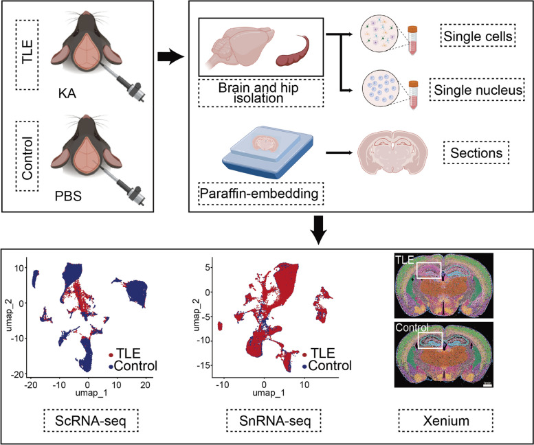
To induce TLE, 200 nl of kainic acid (KA) was stereotactically injected into the hippocampal CA1 region of mice, followed by a 7-day postinjection period. Single-cell RNA sequencing (ScRNA-seq), single-nucleus RNA sequencing (SnRNA-seq), and Xenium-based spatial transcriptomics analyses were employed to evaluate the changes in mRNA expression in glial cells and neurons.
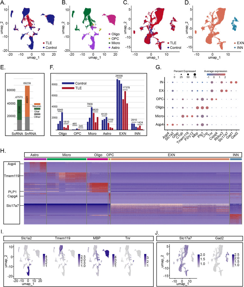
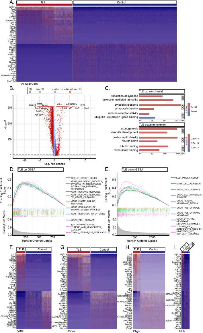
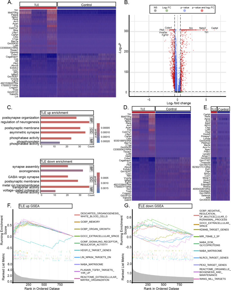
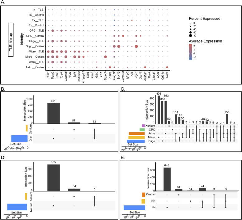
From the ScRNA-seq and SnRNA-seq data, 31,390 glial cells and 48,221 neuronal nuclei were identified. Analysis of the differentially expressed genes (DEGs) revealed significant transcriptomic alterations in the hippocampal cells of mice with TLE, affecting hundreds to thousands of mRNAs and their signalling pathways. Enrichment analysis indicated notable activation of stress and inflammatory pathways in the TLE hippocampus, while pathways related to axonal development and neural support were suppressed. Xenium analysis demonstrated the expression of all 247 genes across mouse brain sections, revealing the spatial distributions of their expression in 27 cell types. Integrated analysis of the DEGs identified via the three sequencing techniques revealed that Spp1, Trem2, and Cd68 were upregulated in all glial cell types and in the Xenium data; Penk, Sorcs3, and Plekha2 were upregulated in all neuronal cell types and in the Xenium data; and Tle4 and Sipa1l3 were downregulated in all glial cell types and in the Xenium data.
In this study, a high-resolution single-cell transcriptomic atlas of the hippocampus in mice with TLE was established, revealing potential intrinsic mechanisms driving TLE-associated inflammatory activation and altered cell interactions. These findings provide valuable insights for further exploration of HS development and epileptogenesis.
颞叶癫痫(TLE)是最常见的癫痫类型之一,频繁发作常导致认知、情感和精神问题。与TLE相关的一个显著病理变化是海马硬化(HS),其特征为神经元丢失、胶质增生和神经元纤维密度增加。TLE-HS发展的潜在机制尚不清楚,但癫痫发生后海马神经胶质细胞和神经元的反应性转录组变化可能提供线索。
为诱导TLE,将200 nl红藻氨酸(KA)立体定向注射到小鼠海马CA1区,注射后观察7天。采用单细胞RNA测序(ScRNA-seq)、单核RNA测序(SnRNA-seq)和基于Xenium的空间转录组学分析来评估神经胶质细胞和神经元中mRNA表达的变化。
从ScRNA-seq和SnRNA-seq数据中,鉴定出31390个神经胶质细胞和48221个神经元细胞核。对差异表达基因(DEG)的分析显示,TLE小鼠海马细胞存在显著的转录组改变,影响数百至数千个mRNA及其信号通路。富集分析表明,TLE海马中应激和炎症通路显著激活,而与轴突发育和神经支持相关的通路受到抑制。Xenium分析显示,所有247个基因在小鼠脑切片中均有表达,揭示了它们在27种细胞类型中的表达空间分布。通过三种测序技术鉴定的DEG的综合分析显示,Spp1、Trem2和Cd68在所有神经胶质细胞类型和Xenium数据中均上调;Penk、Sorcs3和Plekha2在所有神经元细胞类型和Xenium数据中均上调;Tle4和Sipa1l3在所有神经胶质细胞类型和Xenium数据中均下调。
在本研究中,建立了TLE小鼠海马的高分辨率单细胞转录组图谱,揭示了驱动TLE相关炎症激活和细胞相互作用改变的潜在内在机制。这些发现为进一步探索HS发展和癫痫发生提供了有价值的见解。