Higaki Fumiyo, Hiramatsu Masafumi, Yasuhara Takao, Sasada Susumu, Otani Yoshihiro, Haruma Jun, Inoue Tomohiro, Morimitsu Yusuke, Akagi Noriaki, Matsui Yusuke, Iguchi Toshihiro, Hiraki Takao
Department of Radiology, Okayama University Hospital, 2-5-1 Shikata-cho, Kita-ku, Okayama, Japan.
Department of Neurological Surgery, Faculty of Medicine, Dentistry, and Pharmaceutical Sciences, Okayama University, 2-5-1 Shikata-cho, Kita-ku, Okayama, Japan.
Jpn J Radiol. 2025 Feb;43(2):143-151. doi: 10.1007/s11604-024-01661-w. Epub 2024 Sep 16.
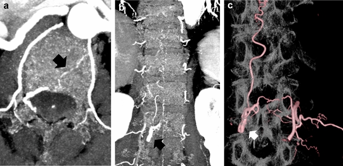
The clinical imaging features of photon-counting detector (PCD) computed tomography (CT) are mainly known as dose reduction, improvement of spatial resolution, and reduction of artifacts compared to energy-integrating detector CT (EID-CT). The utility of cranial and spinal PCD-CT and PCD-CT angiography (CTA) has been previously reported. CTA is a widely used technique for noninvasive evaluation. Cranial CTA is important in brain tumors, especially glioblastoma; it evaluates whether the tumor is highly vascularized prior to an operation and helps in the diagnosis and assessment of bleeding risk. Spinal CTA has an important role in the estimation of feeders and drainers prior to selective angiography in the cases of spinal epidural arteriovenous fistulas and spinal tumors, especially in hemangioblastoma. So far, EID-CTA is commonly performed in an adjunctive role prior to selective angiography; PCD-CTA with high spatial resolution can be an alternative to selective angiography. In the cases of cerebral aneurysms, flow diverters are important tools for the treatment of intracranial aneurysms, and postoperative evaluation with cone beam CT with angiography using diluted contrast media is performed to evaluate stent adhesion and in-stent thrombosis. If CTA can replace selective angiography, it will be less invasive for the patient. In this review, we present representative cases with PCD-CT. We also show how well the cranial and spinal PCD-CTA approaches the accuracy of angiographic and intraoperative findings.
与能量积分探测器CT(EID-CT)相比,光子计数探测器(PCD)计算机断层扫描(CT)的临床成像特征主要表现为辐射剂量降低、空间分辨率提高以及伪影减少。此前已有关于头颅和脊柱PCD-CT及PCD-CT血管造影(CTA)应用的报道。CTA是一种广泛用于无创评估的技术。头颅CTA在脑肿瘤尤其是胶质母细胞瘤中很重要;它在手术前评估肿瘤是否高度血管化,并有助于诊断和评估出血风险。脊柱CTA在脊柱硬膜外动静脉瘘和脊柱肿瘤(尤其是成血管细胞瘤)的选择性血管造影前评估供血动脉和引流静脉方面具有重要作用。到目前为止,EID-CTA通常在选择性血管造影前起辅助作用;具有高空间分辨率的PCD-CTA可以替代选择性血管造影。在脑动脉瘤的病例中,血流导向装置是治疗颅内动脉瘤的重要工具,术后使用稀释造影剂进行锥束CT血管造影评估以评估支架附着情况和支架内血栓形成。如果CTA能够替代选择性血管造影,那么对患者的创伤将更小。在本综述中,我们展示了PCD-CT的典型病例。我们还展示了头颅和脊柱PCD-CTA在接近血管造影和术中发现准确性方面的情况。