Department of Radiology, Mayo Clinic, 200 First Street SW, Rochester, MN, 55905, USA.
Computed Tomography, Siemens Healthineers, Siemensstrasse 3, Forchheim, 91301, Germany.
Eur Radiol. 2023 Aug;33(8):5309-5320. doi: 10.1007/s00330-023-09596-y. Epub 2023 Apr 5.
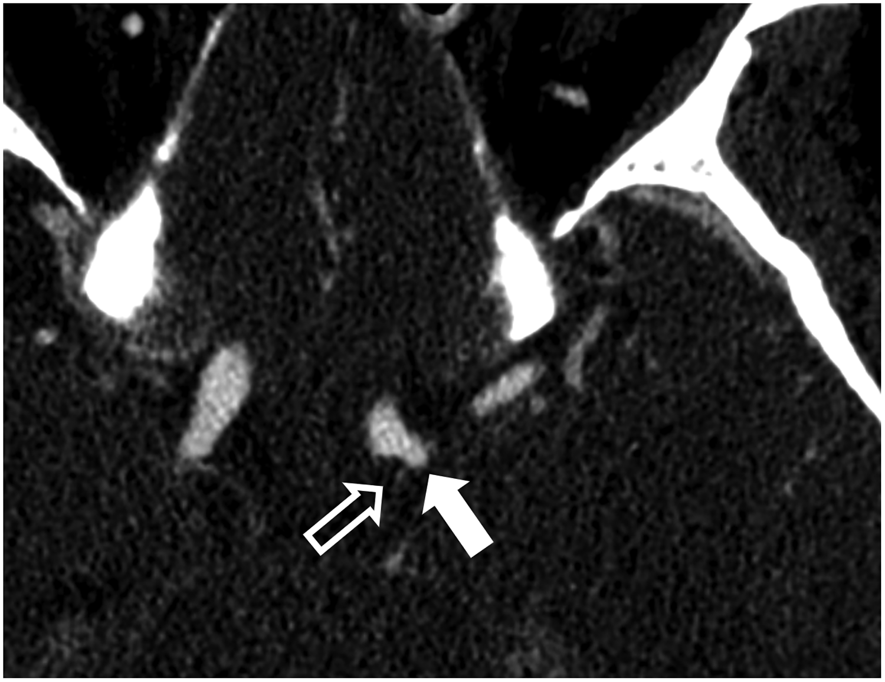
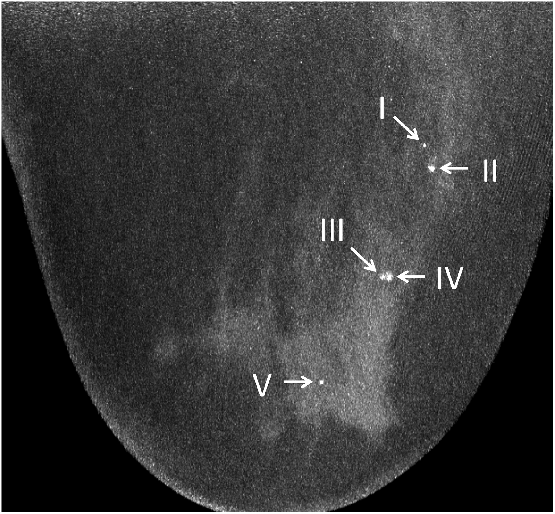
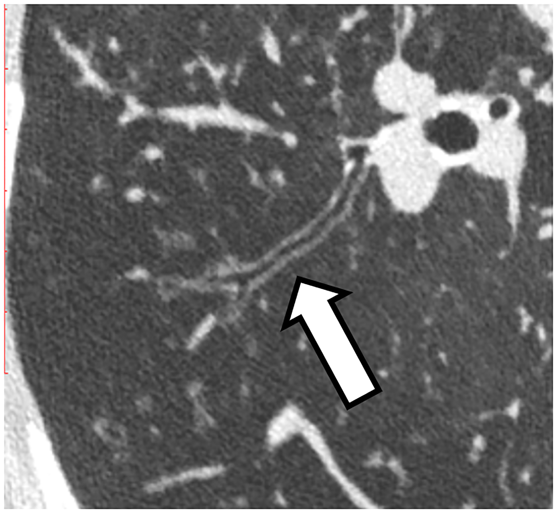
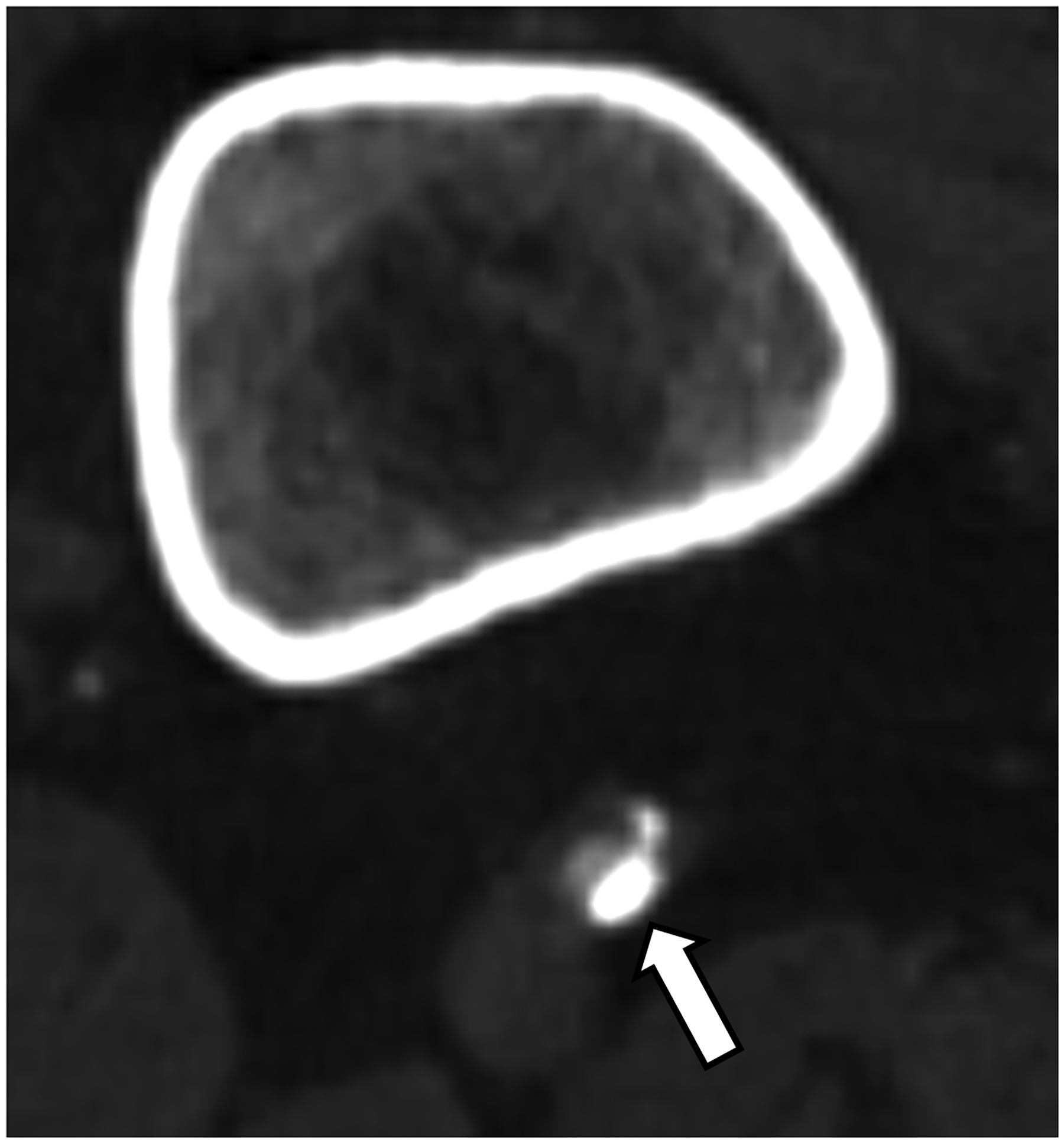
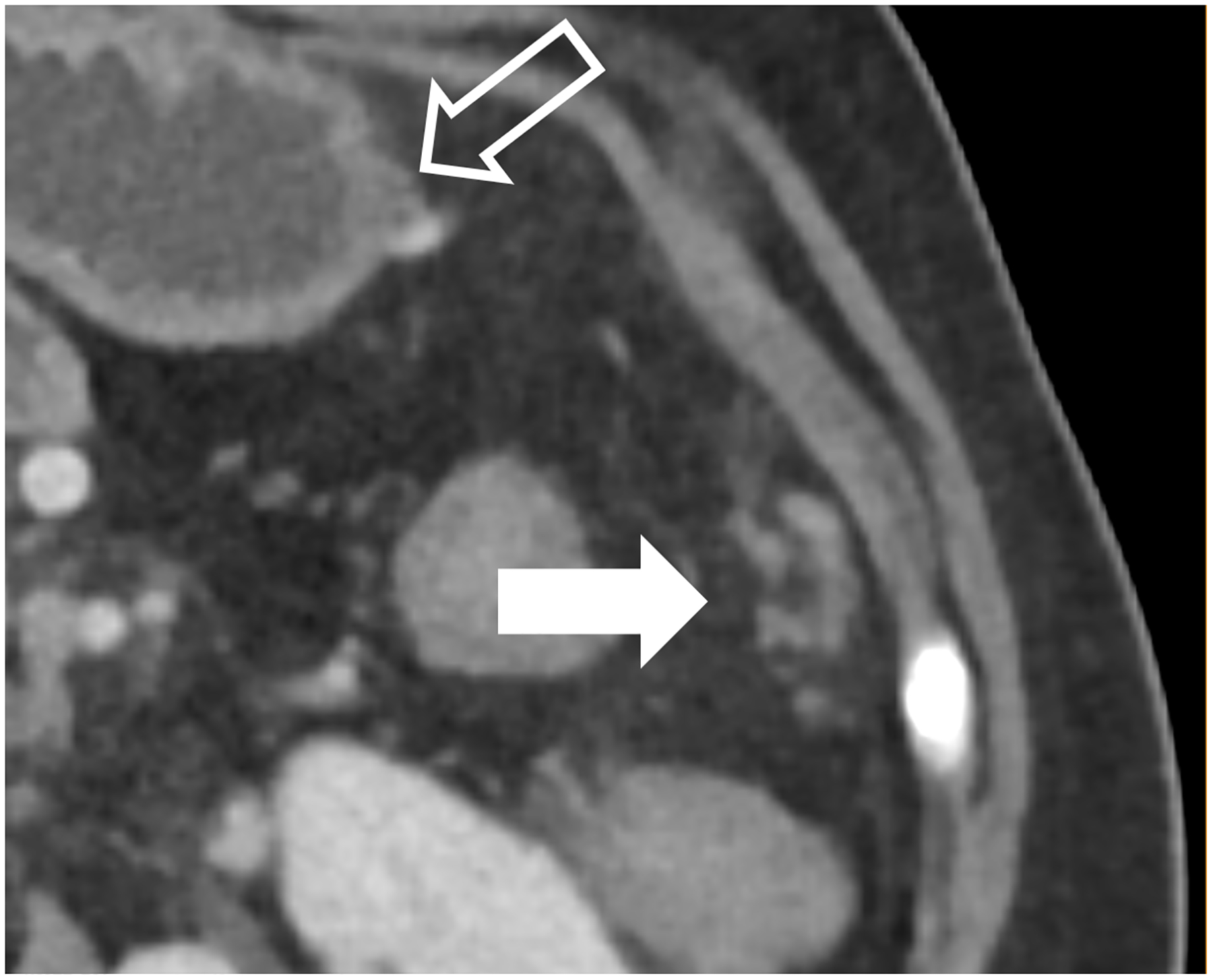
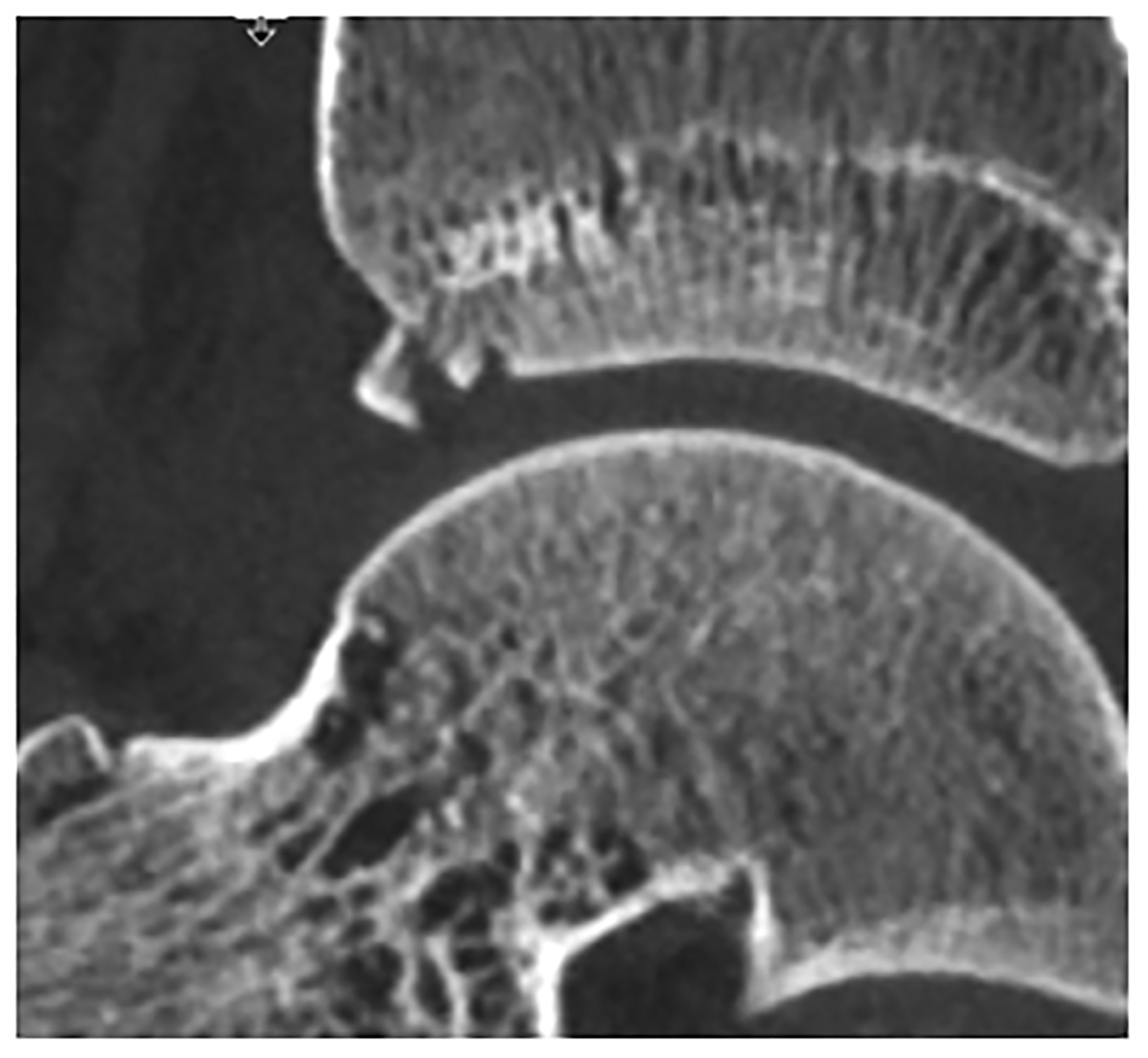
The X-ray detector is a fundamental component of a CT system that determines the image quality and dose efficiency. Until the approval of the first clinical photon-counting-detector (PCD) system in 2021, all clinical CT scanners used scintillating detectors, which do not capture information about individual photons in the two-step detection process. In contrast, PCDs use a one-step process whereby X-ray energy is converted directly into an electrical signal. This preserves information about individual photons such that the numbers of X-ray in different energy ranges can be counted. Primary advantages of PCDs include the absence of electronic noise, improved radiation dose efficiency, increased iodine signal and the ability to use lower doses of iodinated contrast material, and better spatial resolution. PCDs with more than one energy threshold can sort the detected photons into two or more energy bins, making energy-resolved information available for all acquisitions. This allows for material classification or quantitation tasks to be performed in conjunction with high spatial resolution, and in the case of dual-source CT, high pitch, or high temporal resolution acquisitions. Some of the most promising applications of PCD-CT involve imaging of anatomy where exquisite spatial resolution adds clinical value. These include imaging of the inner ear, bones, small blood vessels, heart, and lung. This review describes the clinical benefits observed to date and future directions for this technical advance in CT imaging. KEY POINTS: • Beneficial characteristics of photon-counting detectors include the absence of electronic noise, increased iodine signal-to-noise ratio, improved spatial resolution, and full-time multi-energy imaging. • Promising applications of PCD-CT involve imaging of anatomy where exquisite spatial resolution adds clinical value and applications requiring multi-energy data simultaneous with high spatial and/or temporal resolution. • Future applications of PCD-CT technology may include extremely high spatial resolution tasks, such as the detection of breast micro-calcifications, and quantitative imaging of native tissue types and novel contrast agents.
X 射线探测器是 CT 系统的基本组成部分,决定了图像质量和剂量效率。直到 2021 年第一款临床光子计数探测器(PCD)系统获得批准,所有临床 CT 扫描仪都使用闪烁探测器,在两步检测过程中无法捕捉单个光子的信息。相比之下,PCD 使用一步过程,其中 X 射线能量直接转换为电信号。这保留了关于单个光子的信息,使得可以对不同能量范围内的 X 射线数量进行计数。PCD 的主要优点包括不存在电子噪声、提高辐射剂量效率、增加碘信号和能够使用较低剂量的碘造影剂,以及更好的空间分辨率。具有多个能量阈值的 PCD 可以将检测到的光子分类到两个或更多个能量箱中,从而为所有采集提供能量分辨信息。这允许在结合高空间分辨率的情况下执行物质分类或定量任务,并且在双源 CT 的情况下,高螺距、高时间分辨率采集。PCD-CT 的一些最有前途的应用涉及解剖结构成像,其中精细的空间分辨率增加了临床价值。这些包括内耳、骨骼、小血管、心脏和肺部的成像。这篇综述描述了迄今为止观察到的临床益处以及这项 CT 成像技术进步的未来方向。关键点:
光子计数探测器的有益特征包括不存在电子噪声、增加碘信噪比、提高空间分辨率以及全时多能量成像。
PCD-CT 的有前途的应用涉及解剖结构成像,其中精细的空间分辨率增加了临床价值,以及需要同时具有高空间和/或高时间分辨率的多能量数据的应用。
PCD-CT 技术的未来应用可能包括极高的空间分辨率任务,例如检测乳房微钙化,以及对天然组织类型和新型造影剂的定量成像。