Yunnan Key Laboratory for Basic Research on Bone and Joint Diseases, Kunming University, Kunming, Yunnan, 650214, China.
Yunnan Jici Institute for Regenerative Medicine Co., Ltd, Kunming, Yunnan, 650101, China.
BMC Pulm Med. 2024 Sep 16;24(1):457. doi: 10.1186/s12890-024-03268-3.
Idiopathic pulmonary fibrosis (IPF) is an age-related disease severely affecting life quality with its prevalence rising as the population ages, yet there is still no effective treatment available. Cell therapy has emerged as a promising option for IPF, however, the absence of mature and stable animal models for IPF immunodeficiency hampers preclinical evaluations of human cell therapies, primarily due to rapid immune clearance of administered cells. This study aims to establish a reliable pulmonary fibrosis (PF) model in immunodeficient mice that supports autologous cell therapy and to investigate underlying mechanism.
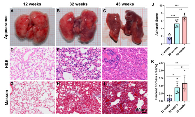
We utilized thirty 5-week-old male NOD/SCID mice, categorizing them into three age groups: 12weeks, 32 weeks and 43 weeks, with 6 mice euthanized randomly from each cohort for lung tissue analysis. We assessed fibrosis using HE staining, Masson's trichrome staining, α-SMA immunohistochemistry and hydroxyproline content measurement. Further, β-galactosidase staining and gene expression analysis of MMP9, TGF-β1, TNF-α, IL-1β, IL-6, IL-8, SOD1, SOD2, NRF2, SIRT1, and SIRT3 were performed. ELISA was employed to quantify protein levels of TNF-α, TGF-β1, and IL-8.
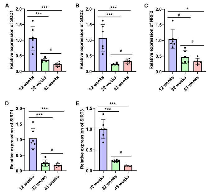
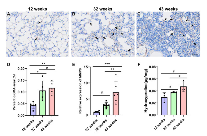
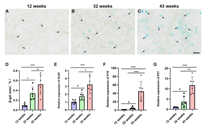
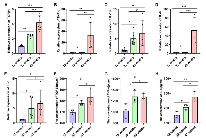
When comparing lung tissues from 32-week-old and 43-week-old mice to those from 12-week-old mice, we noted a marked increase in inflammatory infiltration, fibrosis severity, and hydroxyproline content, alongside elevated expression levels of α-SMA and MMP9. Notably, the degree of fibrosis intensified with age. Additionally, β-galactosidase staining became more pronounced in older mice. Quantitative PCR analyses revealed age-related, increases in the expression of senescence markers (GLB1, P16, P21), and proinflammatory genes (TGF-β1, TNF-α, IL-1β, IL-6, and IL-8). Conversely, the expression of anti-oxidative stress-related genes (SOD1, SOD2, NRF2, SIRT1, and SIRT3) declined, showing statistically significant differences (*P < 0.05, **P < 0.01, ***P < 0.001). ELISA results corroborated these findings, indicating a progressive rise in the protein levels of TGF-β1, TNF-α, and IL-8 as the mice aged.
The findings suggest that NOD/SCID mice aged 32 weeks and 43 weeks effectively model pulmonary fibrosis in an elderly context, with the disease pathogenesis likely driven by age-associated inflammation and oxidative stress.
特发性肺纤维化(IPF)是一种与年龄相关的疾病,严重影响生活质量,随着人口老龄化,其患病率不断上升,但目前尚无有效的治疗方法。细胞疗法已成为 IPF 的一种有前途的选择,然而,缺乏成熟和稳定的用于 IPF 免疫缺陷的动物模型阻碍了人类细胞疗法的临床前评估,主要是因为给予的细胞迅速被免疫清除。本研究旨在建立一种可靠的免疫缺陷小鼠肺纤维化(PF)模型,支持自体细胞治疗,并探讨其潜在机制。
我们使用了 30 只 5 周龄雄性 NOD/SCID 小鼠,将它们分为三组:12 周、32 周和 43 周,每组随机处死 6 只小鼠进行肺组织分析。我们通过 HE 染色、Masson 三色染色、α-SMA 免疫组化和羟脯氨酸含量测定评估纤维化。此外,还进行了β-半乳糖苷酶染色和 MMP9、TGF-β1、TNF-α、IL-1β、IL-6、IL-8、SOD1、SOD2、NRF2、SIRT1 和 SIRT3 的基因表达分析。采用 ELISA 法测定 TNF-α、TGF-β1 和 IL-8 的蛋白水平。
与 12 周龄小鼠的肺组织相比,32 周龄和 43 周龄小鼠的肺组织中炎症浸润、纤维化严重程度和羟脯氨酸含量明显增加,α-SMA 和 MMP9 的表达水平也升高。值得注意的是,纤维化程度随年龄增长而加重。此外,β-半乳糖苷酶染色在老年小鼠中更为明显。定量 PCR 分析显示,衰老相关标志物(GLB1、P16、P21)和促炎基因(TGF-β1、TNF-α、IL-1β、IL-6 和 IL-8)的表达呈年龄相关性增加。相反,抗氧化应激相关基因(SOD1、SOD2、NRF2、SIRT1 和 SIRT3)的表达下降,差异具有统计学意义(*P < 0.05,**P < 0.01,***P < 0.001)。ELISA 结果证实了这些发现,表明随着小鼠年龄的增长,TGF-β1、TNF-α 和 IL-8 的蛋白水平呈逐渐升高趋势。
研究结果表明,32 周龄和 43 周龄的 NOD/SCID 小鼠可有效模拟老年人群中的肺纤维化,疾病发病机制可能与年龄相关的炎症和氧化应激有关。