Department of Gastrointestinal Surgery, Tokyo Medical and Dental University, 1-5-45 Yushima, Bunkyo-ku, Tokyo, Japan.
Department of Gastrointestinal and Pediatric Surgery, Tokyo Medical University, 6-1-1 Shinjuku, Shinjuku-ku, Tokyo, Japan.
BMC Gastroenterol. 2024 Sep 17;24(1):316. doi: 10.1186/s12876-024-03398-2.
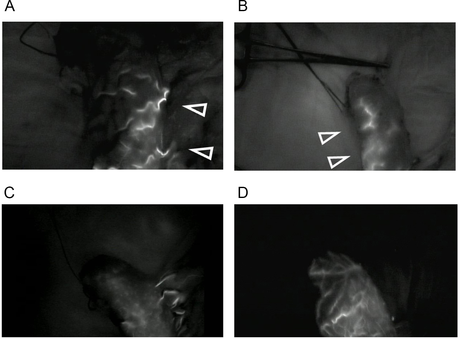
During esophagectomy, evaluation of blood supply to the gastric tube is critically important to estimate and avoid anastomotic complications. This retrospective study investigated the relationship between indocyanine green (ICG) fluorescence angiography during esophagectomy and postoperative endoscopy findings, especially mucosal color change.
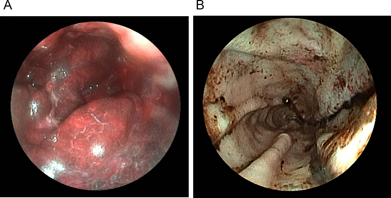
This study retrospectively collected data from 86 patients who underwent subtotal esophagectomy and reconstruction using a gastric tube for esophageal cancer at the Tokyo Medical and Dental University between 2017 and 2020. The flow speed of ICG fluorescence in the gastric tube was evaluated during the operation. Additionally, the main root of ICG enhancement and pattern of ICG distribution in the gastric tube were evaluated. On postoperative day 1 (POD1), the change in the mucosal color to white, thought to reflect ischemia, or black, thought to reflect congestion of the proximal gastric tube, was evaluated. The correlations between these factors, clinical parameters, and surgical outcomes were evaluated. Univariate and multivariate analyses used logistic regression to identify the risk factors affecting mucosal color change.
Multivariate analyses revealed that the only independent significant predictor of mucosal congestion on POD1 was the ICG enhancement time of the right gastric tube tip (odds ratio, 14.49; 95% confidential interval, 2.41-87.24; P = 0.004).
This study indicated that the ICG enhancement time is related to venous malperfusion and congestion rather than arterial malperfusion and ischemia.
在食管切除术期间,评估胃管的血液供应对于估计和避免吻合口并发症至关重要。本回顾性研究调查了食管切除术中吲哚菁绿(ICG)荧光血管造影与术后内镜检查结果之间的关系,特别是黏膜颜色变化。
本研究回顾性收集了 2017 年至 2020 年期间在东京医科齿科大学接受胃管食管癌症根治性切除术的 86 例患者的数据。术中评估胃管中 ICG 荧光的流速。此外,评估了 ICG 增强的主根和胃管中 ICG 分布的模式。术后第 1 天(POD1),评估近端胃管黏膜颜色变白(认为是缺血)或变黑(认为是充血)的变化。评估这些因素与临床参数和手术结果之间的相关性。使用逻辑回归进行单变量和多变量分析,以确定影响黏膜颜色变化的危险因素。
多变量分析显示,POD1 时黏膜充血的唯一独立显著预测因子是胃管右尖端的 ICG 增强时间(比值比,14.49;95%置信区间,2.41-87.24;P = 0.004)。
本研究表明,ICG 增强时间与静脉灌注不良和充血有关,而不是动脉灌注不良和缺血。