• 文献检索
  • 文档翻译
  • 深度研究
  • 学术资讯
  • Suppr Zotero 插件Zotero 插件
  • 邀请有礼
  • 套餐&价格
  • 历史记录
应用&插件
Suppr Zotero 插件Zotero 插件浏览器插件Mac 客户端Windows 客户端微信小程序
定价
高级版会员购买积分包购买API积分包
服务
文献检索文档翻译深度研究API 文档MCP 服务
关于我们
关于 Suppr公司介绍联系我们用户协议隐私条款
关注我们

Suppr 超能文献

核心技术专利:CN118964589B侵权必究
粤ICP备2023148730 号-1Suppr @ 2026

文献检索

告别复杂PubMed语法,用中文像聊天一样搜索,搜遍4000万医学文献。AI智能推荐,让科研检索更轻松。

立即免费搜索

文件翻译

保留排版,准确专业,支持PDF/Word/PPT等文件格式,支持 12+语言互译。

免费翻译文档

深度研究

AI帮你快速写综述,25分钟生成高质量综述,智能提取关键信息,辅助科研写作。

立即免费体验

基于计算机断层扫描影像组学对头颈部癌幸存者下颌骨放射性骨坏死的横断面检测

Computed tomography radiomics-based cross-sectional detection of mandibular osteoradionecrosis in head and neck cancer survivors.

作者信息

Kamel Serageldin, Humbert-Vidan Laia, Kaffey Zaphanlene, Abusaif Abdulrahman, Fuentes David T A, Wahid Kareem, Dede Cem, Naser Mohamed A, He Renjie, Moawad Ahmed W, Elsayes Khaled M, Chen Melissa M, Otun Adegbenga O, Rigert Jillian, Chambers Mark, Hope Andrew, Watson Erin, Brock Kristy K, Hutcheson Katherine, van Dijk Lisanne, Moreno Amy C, Lai Stephen Y, Fuller Clifton D, Mohamed Abdallah S R

机构信息

The University of Texas MD Anderson Cancer Center, Division of Radiation Oncology, Houston, USA.

The University of Texas MD Anderson Cancer Center, Department of Imaging Physics, Houston, USA.

出版信息

medRxiv. 2024 Sep 12:2024.09.11.24313485. doi: 10.1101/2024.09.11.24313485.

DOI:10.1101/2024.09.11.24313485
PMID:39314948
原文链接:https://pmc.ncbi.nlm.nih.gov/articles/PMC11419222/
Abstract

PURPOSE

This study aims to identify radiomic features extracted from contrast-enhanced CT scans that differentiate osteoradionecrosis (ORN) from normal mandibular bone in patients with head and neck cancer (HNC) treated with radiotherapy (RT).

MATERIALS AND METHODS

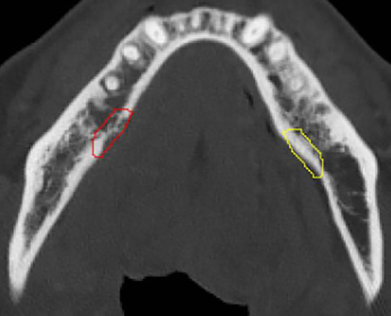
Contrast-enhanced CT (CECT) images were collected for 150 patients (80% train, 20% test) with confirmed ORN diagnosis at The University of Texas MD Anderson Cancer Center between 2008 and 2018. Using PyRadiomics, radiomic features were extracted from manually segmented ORN regions and the corresponding automated control regions, the later defined as the contralateral healthy mandible region. A subset of pre-selected features was obtained based on correlation analysis (r > 0.95) and used to train a Random Forest (RF) classifier with Recursive Feature Elimination. Model explainability SHapley Additive exPlanations (SHAP) analysis was performed on the 20 most important features identified by the trained RF classifier.

RESULTS

From a total of 1316 radiomic features extracted, 810 features were excluded due to high collinearity. From a set of 506 pre-selected radiomic features, the optimal subset resulting on the best discriminative accuracy of the RF classifier consisted of 67 features. The RF classifier was well calibrated (Log Loss 0.296, ECE 0.125) and achieved an accuracy of 88% and a ROC AUC of 0.96. The SHAP analysis revealed that higher values of Wavelet-LLH First-order Mean and Median were associated with ORN of the jaw (ORNJ). Conversely, higher Exponential GLDM Dependence Entropy and lower Square First-order Kurtosis were more characteristic of normal mandibular tissue.

CONCLUSION

This study successfully developed a CECT-based radiomics model for differentiating ORNJ from healthy mandibular tissue in HNC patients after RT. Future work will focus on the detection of subclinical ORNJ regions to guide earlier interventions.

摘要

目的

本研究旨在识别从接受放射治疗(RT)的头颈癌(HNC)患者的对比增强CT扫描中提取的、能区分放射性骨坏死(ORN)与正常下颌骨的放射组学特征。

材料与方法

2008年至2018年期间,在德克萨斯大学MD安德森癌症中心收集了150例确诊为ORN的患者(80%用于训练,20%用于测试)的对比增强CT(CECT)图像。使用PyRadiomics从手动分割的ORN区域和相应的自动对照区域提取放射组学特征,后者定义为对侧健康下颌骨区域。基于相关性分析(r>0.95)获得一组预先选择的特征子集,并用于训练具有递归特征消除的随机森林(RF)分类器。对训练后的RF分类器识别出的20个最重要特征进行模型可解释性SHapley加性解释(SHAP)分析。

结果

在总共提取的1316个放射组学特征中,810个特征因高共线性被排除。从一组506个预先选择的放射组学特征中,使RF分类器具有最佳判别准确性的最优子集由67个特征组成。RF分类器校准良好(对数损失0.296,预期校准误差0.125),准确率达88%,ROC曲线下面积为0.96。SHAP分析表明,小波-LLH一阶均值和中位数的较高值与颌骨放射性骨坏死(ORNJ)相关。相反,指数广义局部二值模式依赖熵较高和平方一阶峰度较低是正常下颌组织更具特征性的表现。

结论

本研究成功开发了一种基于CECT的放射组学模型,用于区分放疗后HNC患者的ORNJ与健康下颌组织。未来的工作将集中在亚临床ORNJ区域的检测上,以指导早期干预。

https://cdn.ncbi.nlm.nih.gov/pmc/blobs/25b3/11419222/7460fa4b660e/nihpp-2024.09.11.24313485v1-f0006.jpg
https://cdn.ncbi.nlm.nih.gov/pmc/blobs/25b3/11419222/efabb58c4022/nihpp-2024.09.11.24313485v1-f0001.jpg
https://cdn.ncbi.nlm.nih.gov/pmc/blobs/25b3/11419222/6e77a7a042e7/nihpp-2024.09.11.24313485v1-f0002.jpg
https://cdn.ncbi.nlm.nih.gov/pmc/blobs/25b3/11419222/ad9421489668/nihpp-2024.09.11.24313485v1-f0003.jpg
https://cdn.ncbi.nlm.nih.gov/pmc/blobs/25b3/11419222/573979ad891a/nihpp-2024.09.11.24313485v1-f0004.jpg
https://cdn.ncbi.nlm.nih.gov/pmc/blobs/25b3/11419222/18b77292f35f/nihpp-2024.09.11.24313485v1-f0005.jpg
https://cdn.ncbi.nlm.nih.gov/pmc/blobs/25b3/11419222/7460fa4b660e/nihpp-2024.09.11.24313485v1-f0006.jpg

相似文献

1
Computed tomography radiomics-based cross-sectional detection of mandibular osteoradionecrosis in head and neck cancer survivors.基于计算机断层扫描影像组学对头颈部癌幸存者下颌骨放射性骨坏死的横断面检测
medRxiv. 2024 Sep 12:2024.09.11.24313485. doi: 10.1101/2024.09.11.24313485.
2
Computed tomography radiomics-based cross-sectional detection of mandibular osteoradionecrosis in head and neck cancer survivors.基于计算机断层扫描影像组学对头颈部癌幸存者下颌骨放射性骨坏死的横断面检测
Oral Oncol. 2025 Aug;167:107337. doi: 10.1016/j.oraloncology.2025.107337. Epub 2025 Jun 13.
3
Machine learning-based radiomics for differentiating lung cancer subtypes in brain metastases using CE-T1WI.基于机器学习的影像组学在使用对比增强T1加权成像鉴别脑转移瘤中肺癌亚型的应用
Front Oncol. 2025 Jun 19;15:1599882. doi: 10.3389/fonc.2025.1599882. eCollection 2025.
4
Integrative radiomics of intra- and peri-tumoral features for enhanced risk prediction in thymic tumors: a multimodal analysis of tumor microenvironment contributions.整合瘤内和瘤周特征的放射组学以增强胸腺瘤风险预测:肿瘤微环境贡献的多模态分析
BMC Med Imaging. 2025 Jul 17;25(1):286. doi: 10.1186/s12880-025-01790-2.
5
Differentiation of Glioblastoma and Solitary Brain Metastasis Using Brain-Tumor Interface Radiomics Features Based on MR Images: A Multicenter Study.基于磁共振图像的脑肿瘤界面放射组学特征鉴别胶质母细胞瘤和孤立性脑转移瘤:一项多中心研究
Acad Radiol. 2025 Jul;32(7):4164-4176. doi: 10.1016/j.acra.2025.04.008. Epub 2025 Apr 24.
6
Comparison of Two Modern Survival Prediction Tools, SORG-MLA and METSSS, in Patients With Symptomatic Long-bone Metastases Who Underwent Local Treatment With Surgery Followed by Radiotherapy and With Radiotherapy Alone.两种现代生存预测工具 SORG-MLA 和 METSSS 在接受手术联合放疗和单纯放疗治疗有症状长骨转移患者中的比较。
Clin Orthop Relat Res. 2024 Dec 1;482(12):2193-2208. doi: 10.1097/CORR.0000000000003185. Epub 2024 Jul 23.
7
Contrast-enhanced ultrasound using SonoVue® (sulphur hexafluoride microbubbles) compared with contrast-enhanced computed tomography and contrast-enhanced magnetic resonance imaging for the characterisation of focal liver lesions and detection of liver metastases: a systematic review and cost-effectiveness analysis.超声造影使用声诺维®(六氟化硫微泡)与对比增强计算机断层扫描和对比增强磁共振成像在局灶性肝脏病变的特征描述和肝转移检测中的比较:系统评价和成本效益分析。
Health Technol Assess. 2013 Apr;17(16):1-243. doi: 10.3310/hta17160.
8
Thymoma habitat segmentation and risk prediction model using CT imaging and K-means clustering.基于CT成像和K均值聚类的胸腺瘤生长部位分割及风险预测模型
Med Phys. 2025 Jul;52(7):e17892. doi: 10.1002/mp.17892. Epub 2025 May 19.
9
Prediction of KRAS gene mutations in colorectal cancer using a CT-based radiomic model.基于CT的影像组学模型预测结直肠癌中的KRAS基因突变
Front Med (Lausanne). 2025 May 12;12:1592497. doi: 10.3389/fmed.2025.1592497. eCollection 2025.
10
The impact of uncertainty estimation on radiomic segmentation reproducibility and scan-rescan repeatability in kidney MRI.不确定性估计对肾脏MRI中放射组学分割再现性和扫描-重扫重复性的影响。
Med Phys. 2025 Jul;52(7):e17995. doi: 10.1002/mp.17995.

本文引用的文献

1
Prevention and Management of Osteoradionecrosis in Patients With Head and Neck Cancer Treated With Radiation Therapy: ISOO-MASCC-ASCO Guideline.头颈部癌症放射治疗患者的放射性骨坏死的预防和管理:ISOO-MASCC-ASCO 指南。
J Clin Oncol. 2024 Jun 1;42(16):1975-1996. doi: 10.1200/JCO.23.02750. Epub 2024 May 1.
2
Computed Tomography Radiomics Kinetics as Early Imaging Correlates of Osteoradionecrosis in Oropharyngeal Cancer Patients.计算机断层扫描放射组学动力学作为口咽癌患者放射性骨坏死的早期影像学关联指标
Front Artif Intell. 2021 Apr 9;4:618469. doi: 10.3389/frai.2021.618469. eCollection 2021.
3
The Image Biomarker Standardization Initiative: Standardized Quantitative Radiomics for High-Throughput Image-based Phenotyping.
影像生物标志物标准化倡议:高通量基于影像表型的标准化定量放射组学。
Radiology. 2020 May;295(2):328-338. doi: 10.1148/radiol.2020191145. Epub 2020 Mar 10.
4
A Radiomics Nomogram for Preoperative Prediction of Microvascular Invasion in Hepatocellular Carcinoma.一种用于术前预测肝细胞癌微血管侵犯的影像组学列线图。
Liver Cancer. 2019 Oct;8(5):373-386. doi: 10.1159/000494099. Epub 2018 Nov 27.
5
Radiomics signature extracted from diffusion-weighted magnetic resonance imaging predicts outcomes in osteosarcoma.从扩散加权磁共振成像中提取的影像组学特征可预测骨肉瘤的预后。
J Bone Oncol. 2019 Oct 4;19:100263. doi: 10.1016/j.jbo.2019.100263. eCollection 2019 Dec.
6
The prognostic value of CT-based image-biomarkers for head and neck cancer patients treated with definitive (chemo-)radiation.基于 CT 的影像学生物标志物对头颈部癌症患者接受根治性(放化疗)治疗的预后价值。
Oral Oncol. 2019 Aug;95:178-186. doi: 10.1016/j.oraloncology.2019.06.020. Epub 2019 Jun 26.
7
Radiomics in Glioblastoma: Current Status and Challenges Facing Clinical Implementation.胶质母细胞瘤中的放射组学:临床应用现状与面临的挑战
Front Oncol. 2019 May 21;9:374. doi: 10.3389/fonc.2019.00374. eCollection 2019.
8
Novel imaging techniques of rectal cancer: what do radiomics and radiogenomics have to offer? A literature review.直肠癌的新型影像学技术:放射组学和放射基因组学有何贡献?文献综述。
Abdom Radiol (NY). 2019 Nov;44(11):3764-3774. doi: 10.1007/s00261-019-02042-y.
9
Magnetic resonance imaging (MRI)-based radiomics for prostate cancer radiotherapy.基于磁共振成像(MRI)的放射组学在前列腺癌放疗中的应用
Transl Androl Urol. 2018 Jun;7(3):445-458. doi: 10.21037/tau.2018.06.05.
10
Radiomics in gliomas: A promising assistance 
for glioma clinical research.胶质瘤中的放射组学:对胶质瘤临床研究的一项有前景的辅助手段。
Zhong Nan Da Xue Xue Bao Yi Xue Ban. 2018 Apr 28;43(4):354-359. doi: 10.11817/j.issn.1672-7347.2018.04.004.