• 文献检索
  • 文档翻译
  • 深度研究
  • 学术资讯
  • Suppr Zotero 插件Zotero 插件
  • 邀请有礼
  • 套餐&价格
  • 历史记录
应用&插件
Suppr Zotero 插件Zotero 插件浏览器插件Mac 客户端Windows 客户端微信小程序
定价
高级版会员购买积分包购买API积分包
服务
文献检索文档翻译深度研究API 文档MCP 服务
关于我们
关于 Suppr公司介绍联系我们用户协议隐私条款
关注我们

Suppr 超能文献

核心技术专利:CN118964589B侵权必究
粤ICP备2023148730 号-1Suppr @ 2026

文献检索

告别复杂PubMed语法,用中文像聊天一样搜索,搜遍4000万医学文献。AI智能推荐,让科研检索更轻松。

立即免费搜索

文件翻译

保留排版,准确专业,支持PDF/Word/PPT等文件格式,支持 12+语言互译。

免费翻译文档

深度研究

AI帮你快速写综述,25分钟生成高质量综述,智能提取关键信息,辅助科研写作。

立即免费体验

房间隔脂肪性肥厚:慢性阻塞性肺疾病中一种独特的脂肪组织类型?

Lipomatous hypertrophy of the interatrial septum: a distinct adipose tissue type in COPD?

作者信息

Lacaita Pietro G, Kindl Benedikt, Plank Fabian, Beyer Christoph, Bilgeri Valentin, Barbieri Fabian, Senoner Thomas, Dichtl Wolfgang, Tancevski Ivan, Swoboda Michael, Luger Anna, Deeg Johannes, Widmann Gerlig, Feuchtner Gudrun M

机构信息

Department of Radiology, Innsbruck Medical University, Innsbruck, Austria.

Department of Internal Medicine, Tyrol Clinicum Hall, Hall, Austria.

出版信息

ERJ Open Res. 2024 Sep 23;10(5). doi: 10.1183/23120541.00295-2024. eCollection 2024 Sep.

DOI:10.1183/23120541.00295-2024
PMID:39319042
原文链接:https://pmc.ncbi.nlm.nih.gov/articles/PMC11417605/
Abstract

OBJECTIVE

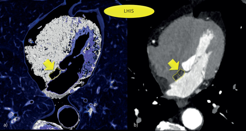
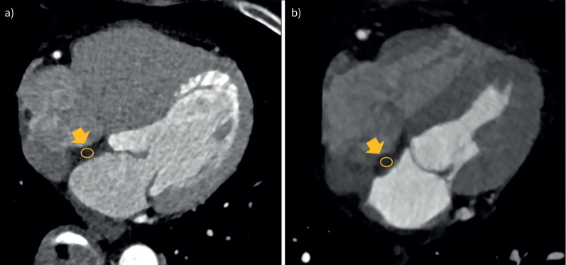
Lipomatous hypertrophy of the interatrial septum (LHIS) is a distinct section of epicardial adipose tissue. However, its association with COPD is poorly documented.

METHODS

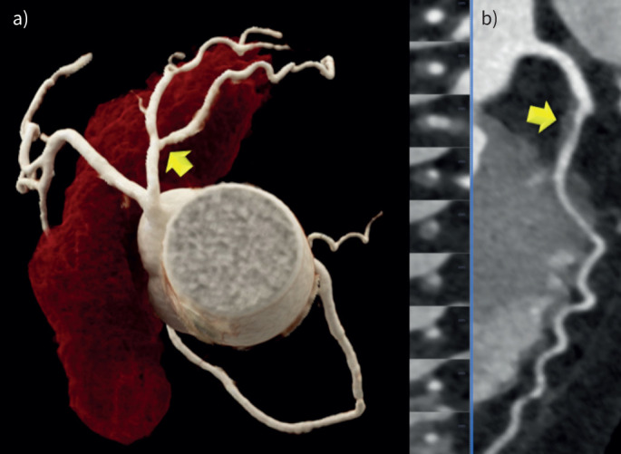
Patients undergoing coronary computed tomography angiography (CTA) for clinical indications were recruited retrospectively and screened for LHIS and COPD. LHIS density and the coronary artery disease profile were quantified by CTA: stenosis severity (coronary artery disease radiological reporting system (CADRADS)), coronary artery calcium (CAC) and high-risk plaque (HRP). COPD patients with LHIS were matched for age and sex, the major cardiovascular risk factors (CVRFs), and compared to controls.

RESULTS

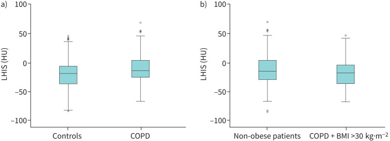
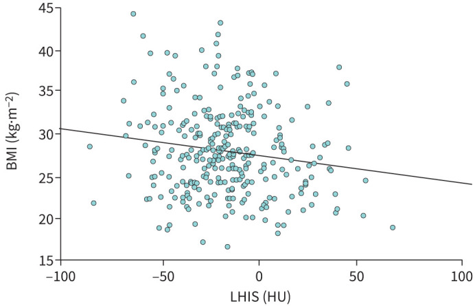
The prevalence of LHIS in all 5466 patients was 5.9%. 151 (72.6%) of 208 patients with COPD had LHIS. LHIS density in COPD patients was higher (-10.93 HU -21.1 HU; p<0.001), despite body mass index (BMI) (28.8 27.01 kg·m; p=0.002) being lower. LHIS density was lower in obese (BMI >30 kg·m) patients (20.4 13.6 HU; p=0.02). BMI was inversely correlated with LHIS density (BetaR -0.031; 95% CI: -0.054- -0.008; p=0.007). LHIS density was associated with COPD, but not with BMI on multivariate models. CAC and coronary stenosis severity (CADRADS and >50% stenosis) were not different (p=0.106, p=0.156 and p=0.350, respectively). HRPs were observed more frequently in COPD patients with severe Global Initiative for Chronic Obstructive Lung Disease (GOLD) stages ≥2 (32.3% 20.1%; p=0.044), but not when adding mild GOLD stages.

CONCLUSIONS

The prevalence of LHIS in COPD patients is high (72.6%), and the adipose tissue density is higher, indicating a higher brown fat component. In obese, patients LHIS density is lower and declines along with BMI. Coronary stenosis severity and calcium were not different; however HRPs were more frequent in severe COPD.

摘要

目的

房间隔脂肪肥厚(LHIS)是心外膜脂肪组织的一个独特部分。然而,其与慢性阻塞性肺疾病(COPD)的关联鲜有文献记载。

方法

回顾性招募因临床指征接受冠状动脉计算机断层扫描血管造影(CTA)的患者,筛查LHIS和COPD。通过CTA对LHIS密度和冠状动脉疾病特征进行量化:狭窄严重程度(冠状动脉疾病放射学报告系统(CADRADS))、冠状动脉钙化(CAC)和高危斑块(HRP)。将患有LHIS的COPD患者按年龄、性别、主要心血管危险因素(CVRFs)进行匹配,并与对照组进行比较。

结果

在所有5466例患者中,LHIS的患病率为5.9%。208例COPD患者中有151例(72.6%)患有LHIS。尽管体重指数(BMI)较低(28.8对27.01kg·m²;p=0.002),但COPD患者的LHIS密度更高(-10.93HU对-21.1HU;p<0.001)。肥胖(BMI>30kg·m²)患者的LHIS密度较低(20.4对13.6HU;p=0.02)。BMI与LHIS密度呈负相关(βR -0.031;95%CI:-0.054至-0.008;p=0.007)。在多变量模型中,LHIS密度与COPD相关,但与BMI无关。CAC和冠状动脉狭窄严重程度(CADRADS和>50%狭窄)无差异(分别为p=0.106、p=0.156和p=0.350)。在慢性阻塞性肺疾病全球倡议(GOLD)≥2期的重度COPD患者中,HRP的观察频率更高(32.3%对20.1%;p=0.044),但加入轻度GOLD期时则不然。

结论

COPD患者中LHIS的患病率较高(72.6%),且脂肪组织密度更高,表明棕色脂肪成分更多。在肥胖患者中,LHIS密度较低且随BMI下降。冠状动脉狭窄严重程度和钙化无差异;然而,重度COPD患者中HRP更常见。

https://cdn.ncbi.nlm.nih.gov/pmc/blobs/d429/11417605/875df4b0e1bc/00295-2024.05.jpg
https://cdn.ncbi.nlm.nih.gov/pmc/blobs/d429/11417605/47d9ff2572ef/00295-2024.01.jpg
https://cdn.ncbi.nlm.nih.gov/pmc/blobs/d429/11417605/bfb5b57fcc5e/00295-2024.02.jpg
https://cdn.ncbi.nlm.nih.gov/pmc/blobs/d429/11417605/b96c3b834c40/00295-2024.03.jpg
https://cdn.ncbi.nlm.nih.gov/pmc/blobs/d429/11417605/feec0abba039/00295-2024.04.jpg
https://cdn.ncbi.nlm.nih.gov/pmc/blobs/d429/11417605/875df4b0e1bc/00295-2024.05.jpg

相似文献

1
Lipomatous hypertrophy of the interatrial septum: a distinct adipose tissue type in COPD?房间隔脂肪性肥厚:慢性阻塞性肺疾病中一种独特的脂肪组织类型?
ERJ Open Res. 2024 Sep 23;10(5). doi: 10.1183/23120541.00295-2024. eCollection 2024 Sep.
2
The interaction of lipomatous hypertrophy of the interatrial septum with pericardial adipose tissue biomarkers by computed tomography.通过计算机断层扫描研究房间隔脂肪肥厚与心包脂肪组织生物标志物的相互作用。
Eur Radiol. 2025 Apr;35(4):2189-2199. doi: 10.1007/s00330-024-11061-3. Epub 2024 Sep 5.
3
Lipomatous hypertrophy of the interatrial septum: prevalence and features on fusion 18F fluorodeoxyglucose positron emission tomography/CT.房间隔脂肪肥厚:18F氟脱氧葡萄糖正电子发射断层扫描/计算机断层扫描融合图像上的患病率及特征
Chest. 2005 Dec;128(6):3888-93. doi: 10.1378/chest.128.6.3888.
4
Visceral adiposity in patients with lipomatous hypertrophy of the interatrial septum.脂肪心在房间隔脂肪肥厚患者中的表现。
Heart Vessels. 2024 Feb;39(2):160-166. doi: 10.1007/s00380-023-02319-9. Epub 2023 Oct 4.
5
Lipomatous hypertrophy of the interatrial septum in ECG-gated multislice computed tomography of the heart.心脏心电门控多层螺旋 CT 中的房间隔脂肪性肥厚。
Med Sci Monit. 2012 Jul;18(7):MT54-9. doi: 10.12659/msm.883197.
6
Peri-Coronary Adipose Tissue Density Is Associated With F-Sodium Fluoride Coronary Uptake in Stable Patients With High-Risk Plaques.冠状动脉周围脂肪组织密度与高危斑块稳定患者 F-氟脱氧葡萄糖冠状动脉摄取相关。
JACC Cardiovasc Imaging. 2019 Oct;12(10):2000-2010. doi: 10.1016/j.jcmg.2018.11.032. Epub 2019 Feb 13.
7
A case report of "lipomatous hypertrophy of the cardiac interatrial septum", with a proposal for a new term "lipomatous hamartoma of the cardiac atrial septum".一例“心脏房间隔脂肪性肥厚”病例报告,并提议采用新术语“心脏房间隔脂肪性错构瘤”
Acta Pathol Jpn. 1988 Dec;38(12):1583-9. doi: 10.1111/j.1440-1827.1988.tb02298.x.
8
FDG uptake in lipomatous hypertrophy of the interatrial septum is not likely related to brown adipose tissue.房间隔脂肪肥厚中 FDG 的摄取与棕色脂肪组织无关。
Clin Nucl Med. 2011 Sep;36(9):767-9. doi: 10.1097/RLU.0b013e318219b353.
9
Lipomatous hypertrophy of the interatrial septum: in vivo diagnosis.房间隔脂肪肥厚:活体诊断
Circulation. 1982 Aug;66(2):470-3. doi: 10.1161/01.cir.66.2.470.
10
Persistent high grade flurodeoxyglucose uptake in lipomatous hypertrophy of the interatrial septum on dual time point imaging and with ambient warming.在双时相成像及环境升温情况下,房间隔脂肪性肥厚部位持续存在高等级氟脱氧葡萄糖摄取。
World J Nucl Med. 2014 Jan;13(1):62-3. doi: 10.4103/1450-1147.138577.

本文引用的文献

1
Cardiovascular Significance and Genetics of Epicardial and Pericardial Adiposity.心外膜和心包脂肪的心血管意义和遗传学。
JAMA Cardiol. 2024 May 1;9(5):418-427. doi: 10.1001/jamacardio.2024.0080.
2
Epicardial adipose tissue in patients with chronic obstructive pulmonary disease: systematic review with meta‑analysis and trial sequential analysis.慢性阻塞性肺疾病患者的心外膜脂肪组织:系统评价与荟萃分析及试验序贯分析。
BMC Pulm Med. 2023 Jul 3;23(1):241. doi: 10.1186/s12890-023-02535-z.
3
GOLD COPD DOCUMENT 2023: a brief update for practicing cardiologists.
2023 年 GOLD COPD 文档:对执业心脏病学家的简要更新。
Clin Res Cardiol. 2024 Feb;113(2):195-204. doi: 10.1007/s00392-023-02217-0. Epub 2023 May 26.
4
Global Initiative for Chronic Obstructive Lung Disease 2023 Report: GOLD Executive Summary.慢性阻塞性肺疾病全球倡议组织2023年报告:《慢性阻塞性肺疾病全球倡议》执行摘要
Am J Respir Crit Care Med. 2023 Apr 1;207(7):819-837. doi: 10.1164/rccm.202301-0106PP.
5
Hypoxia as a Double-Edged Sword to Combat Obesity and Comorbidities.缺氧作为对抗肥胖及其合并症的双刃剑。
Cells. 2022 Nov 23;11(23):3735. doi: 10.3390/cells11233735.
6
Atrial fibrillation and chronic obstructive pulmonary disease: diagnostic sequence and mortality risk.心房颤动与慢性阻塞性肺疾病:诊断顺序及死亡风险
Eur Heart J Qual Care Clin Outcomes. 2023 Feb 28;9(2):128-134. doi: 10.1093/ehjqcco/qcac059.
7
Factors associated with coronary heart disease in COPD patients and controls.COPD 患者和对照者中与冠心病相关的因素。
PLoS One. 2022 Apr 27;17(4):e0265682. doi: 10.1371/journal.pone.0265682. eCollection 2022.
8
The Atherosclerotic Profile of a Young Symptomatic Population between 19 and 49 Years: Coronary Computed Tomography Angiography or Coronary Artery Calcium Score?19至49岁有症状年轻人群的动脉粥样硬化特征:冠状动脉计算机断层扫描血管造影还是冠状动脉钙化积分?
J Cardiovasc Dev Dis. 2021 Nov 18;8(11):157. doi: 10.3390/jcdd8110157.
9
Reply: Prognostic Role of Pericardial Fat on the Incidence of Heart Failure.回复:心包脂肪对心力衰竭发生率的预后作用。
J Am Coll Cardiol. 2021 Oct 12;78(15):e113-e115. doi: 10.1016/j.jacc.2021.08.008.
10
Prevalence, management and impact of chronic obstructive pulmonary disease in atrial fibrillation: a systematic review and meta-analysis of 4,200,000 patients.心房颤动患者中慢性阻塞性肺疾病的患病率、管理及影响:对420万例患者的系统评价和荟萃分析
Eur Heart J. 2021 Sep 14;42(35):3541-3554. doi: 10.1093/eurheartj/ehab453.