• 文献检索
  • 文档翻译
  • 深度研究
  • 学术资讯
  • Suppr Zotero 插件Zotero 插件
  • 邀请有礼
  • 套餐&价格
  • 历史记录
应用&插件
Suppr Zotero 插件Zotero 插件浏览器插件Mac 客户端Windows 客户端微信小程序
定价
高级版会员购买积分包购买API积分包
服务
文献检索文档翻译深度研究API 文档MCP 服务
关于我们
关于 Suppr公司介绍联系我们用户协议隐私条款
关注我们

Suppr 超能文献

核心技术专利:CN118964589B侵权必究
粤ICP备2023148730 号-1Suppr @ 2026

文献检索

告别复杂PubMed语法,用中文像聊天一样搜索,搜遍4000万医学文献。AI智能推荐,让科研检索更轻松。

立即免费搜索

文件翻译

保留排版,准确专业,支持PDF/Word/PPT等文件格式,支持 12+语言互译。

免费翻译文档

深度研究

AI帮你快速写综述,25分钟生成高质量综述,智能提取关键信息,辅助科研写作。

立即免费体验

大鼠(大鼠胸部超声):宠物大鼠气胸的诊断

RATTUS (Rat Thoracic Ultrasound): diagnosis of pneumothorax in pet rats.

作者信息

Piskovská A, Kraszewska K, Hauptman K, Chloupek J, Linhart P, Jekl V

机构信息

Jekl & Hauptman Veterinary Clinic, Brno, Czechia.

Department of Pharmacology and Pharmacy, Faculty of Veterinary Medicine, VETUNI, Brno, Czechia.

出版信息

Front Vet Sci. 2024 Sep 13;11:1394291. doi: 10.3389/fvets.2024.1394291. eCollection 2024.

DOI:10.3389/fvets.2024.1394291
PMID:39346960
原文链接:https://pmc.ncbi.nlm.nih.gov/articles/PMC11428198/
Abstract

INTRODUCTION

Rat thoracic ultrasound (RATTUS) is a non-invasive, easy-to-perform method for the evaluation of the pleural space and lungs in pet rats. The aim of the article is to present species-specific differences in the sonographic diagnosis of pneumothorax (PTX) in pet rats.

METHODS

In total, 158 client-owned pet rats were examined during the period from July 2023 to January 2024. PTX was diagnosed in 20 of the examined rats (13.25%, the age of the animals ranged from 2 months to 32 months (19.08 ± 6.93 months; mean ± SD) and their body weight ranged from 97 g to 885 g (461.27 ± 138.97 g; mean ± SD). Radiographic confirmation of PTX was performed in all these 20 rats, in the control group radiography was used to confirm that PTX was not present.

RESULTS

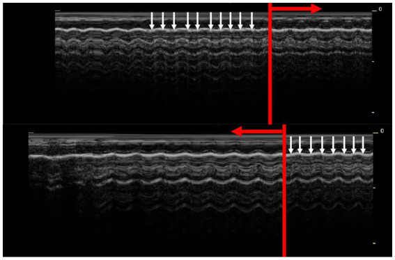
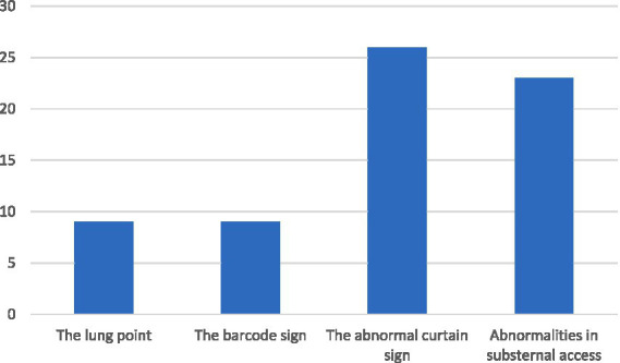
The lung point and the barcode sign was found in 7/20 animals with sensitivity of 33.3% (95% CI, 0.16-0.59) and specificity of 100% (95% CI, 0.97-1.0). The abnormal curtain sign was found in 19/20 of animals with the sensitivity of 95% (95% CI, 0.73-0.99.7) and the specificity of 89% (95% CI, 0.82-0.93). The abnormalities in the substernal access were in 17/20 of animals with the sensitivity of 85% (95% CI, 0.61-0.96) and the specificity of 71% (95% CI, 0.62-0.78).

DISCUSSION

In conclusion, RATTUS is a non-invasive method for the diagnosis of PTX in rats. Lung point and barcode sign are specific but not easily diagnosed signs. The curtain sign in RATTUS is not specific for PTX, as there are e.g. geriatric rats (rats older than 1,5 years) in which the abnormal curtain sign is visible without the presence of PTX. The presence of moderate to severe PTX can be assessed by the substernal approach based on the presence of cardiac displacement toward the collapsed lung lobe, and on evaluation of the lung inflation symmetry. This sign is not specific for PTX but in conjunction with other ultrasonic signs described makes the RATTUS a feasible tool for PTX diagnosis in rats.

摘要

引言

大鼠胸部超声检查(RATTUS)是一种用于评估宠物大鼠胸膜腔和肺部的非侵入性、易于操作的方法。本文旨在介绍宠物大鼠气胸(PTX)超声诊断中的物种特异性差异。

方法

在2023年7月至2024年1月期间,共检查了158只客户拥有的宠物大鼠。在检查的大鼠中有20只被诊断为气胸(13.25%),这些动物的年龄在2个月至32个月之间(19.08±6.93个月;平均值±标准差),体重在97克至885克之间(461.27±138.97克;平均值±标准差)。对所有这20只大鼠进行了气胸的X线确认,在对照组中,使用X线检查确认不存在气胸。

结果

在20只动物中有7只发现了肺点和条形码征,敏感性为33.3%(95%可信区间,0.16 - 0.59),特异性为100%(95%可信区间,0.97 - 1.0)。在20只动物中有19只发现了异常帷幕征,敏感性为95%(95%可信区间,0.73 - 0.997),特异性为89%(95%可信区间,0.82 - 0.93)。在20只动物中有17只发现胸骨下通路异常,敏感性为85%(95%可信区间,0.61 - 0.96),特异性为71%(95%可信区间,0.62 - 0.78)。

讨论

总之,RATTUS是一种用于诊断大鼠气胸的非侵入性方法。肺点和条形码征具有特异性,但不易诊断。RATTUS中的帷幕征并非气胸所特有,例如在老年大鼠(年龄超过1.5岁)中,即使没有气胸也可见异常帷幕征。中度至重度气胸的存在可通过胸骨下途径评估,基于心脏向塌陷肺叶的移位以及肺膨胀对称性的评估。该征象并非气胸所特有,但与所描述的其他超声征象结合,使RATTUS成为诊断大鼠气胸的可行工具。

https://cdn.ncbi.nlm.nih.gov/pmc/blobs/93a3/11428198/adc6ff883fe0/fvets-11-1394291-g003.jpg
https://cdn.ncbi.nlm.nih.gov/pmc/blobs/93a3/11428198/f8d0d11f3aee/fvets-11-1394291-g001.jpg
https://cdn.ncbi.nlm.nih.gov/pmc/blobs/93a3/11428198/08d7d192fb8b/fvets-11-1394291-g002.jpg
https://cdn.ncbi.nlm.nih.gov/pmc/blobs/93a3/11428198/adc6ff883fe0/fvets-11-1394291-g003.jpg
https://cdn.ncbi.nlm.nih.gov/pmc/blobs/93a3/11428198/f8d0d11f3aee/fvets-11-1394291-g001.jpg
https://cdn.ncbi.nlm.nih.gov/pmc/blobs/93a3/11428198/08d7d192fb8b/fvets-11-1394291-g002.jpg
https://cdn.ncbi.nlm.nih.gov/pmc/blobs/93a3/11428198/adc6ff883fe0/fvets-11-1394291-g003.jpg

相似文献

1
RATTUS (Rat Thoracic Ultrasound): diagnosis of pneumothorax in pet rats.大鼠(大鼠胸部超声):宠物大鼠气胸的诊断
Front Vet Sci. 2024 Sep 13;11:1394291. doi: 10.3389/fvets.2024.1394291. eCollection 2024.
2
The Rat Thoracic Ultrasound protocol: scanning technique and normal findings.大鼠胸部超声检查方案:扫描技术及正常表现
Front Vet Sci. 2024 Feb 19;11:1286614. doi: 10.3389/fvets.2024.1286614. eCollection 2024.
3
Abnormal Curtain Signs Identified With a Novel Lung Ultrasound Protocol in Six Dogs With Pneumothorax.采用新型肺部超声方案在六只气胸犬中识别出异常帷幕征。
Front Vet Sci. 2019 Aug 28;6:291. doi: 10.3389/fvets.2019.00291. eCollection 2019.
4
A prospective comparison of supine chest radiography and bedside ultrasound for the diagnosis of traumatic pneumothorax.仰卧位胸部X线摄影与床边超声诊断创伤性气胸的前瞻性比较
Acad Emerg Med. 2005 Sep;12(9):844-9. doi: 10.1197/j.aem.2005.05.005.
5
Lung Ultrasonography for Pneumothorax in Dogs and Cats.犬猫气胸的肺部超声检查
Vet Clin North Am Small Anim Pract. 2021 Nov;51(6):1153-1167. doi: 10.1016/j.cvsm.2021.07.003. Epub 2021 Sep 9.
6
Lung Ultrasound Diagnostic Accuracy in Neonatal Pneumothorax.肺部超声对新生儿气胸的诊断准确性
Can Respir J. 2016;2016:6515069. doi: 10.1155/2016/6515069. Epub 2016 May 3.
7
Computerized Diagnostic Assistant for the Automatic Detection of Pneumothorax on Ultrasound: A Pilot Study.用于超声自动检测气胸的计算机诊断助手:一项初步研究。
West J Emerg Med. 2016 Mar;17(2):209-15. doi: 10.5811/westjem.2016.1.28087. Epub 2016 Mar 2.
8
Accuracy of Resident-Performed Point-of-Care Lung Ultrasound Examinations Versus Chest Radiography in Pneumothorax Follow-up After Tube Thoracostomy in Rwanda.卢旺达经胸管引流术后气胸随访中,由住院医师行床边肺部超声检查与胸部 X 线检查的准确性比较。
J Ultrasound Med. 2020 Mar;39(3):499-506. doi: 10.1002/jum.15126. Epub 2019 Sep 6.
9
Role of ultrasonography in the diagnosis and management of pneumothorax following transbronchial lung biopsy.超声检查在经支气管肺活检术后气胸诊断及处理中的作用
J Bronchology Interv Pulmonol. 2015 Jan;22(1):14-9. doi: 10.1097/LBR.0000000000000125.
10
Diagnostic value of pulsed wave doppler in pneumothorax: a prospective study.脉冲多普勒在气胸诊断中的价值:一项前瞻性研究。
Ir J Med Sci. 2024 Apr;193(2):1025-1031. doi: 10.1007/s11845-023-03513-4. Epub 2023 Sep 7.

本文引用的文献

1
Retrospective evaluation of the agreement between thoracic point-of-care ultrasound and thoracic radiographs in cats with recent trauma: 111 cats.对近期遭受创伤的猫进行胸部床旁超声与胸部X光片一致性的回顾性评估:111只猫。
Front Vet Sci. 2024 Jun 26;11:1376004. doi: 10.3389/fvets.2024.1376004. eCollection 2024.
2
The Rat Thoracic Ultrasound protocol: scanning technique and normal findings.大鼠胸部超声检查方案:扫描技术及正常表现
Front Vet Sci. 2024 Feb 19;11:1286614. doi: 10.3389/fvets.2024.1286614. eCollection 2024.
3
Case report: Application of color Doppler sonography for the assessment of pulmonary consolidations in a dog.
病例报告:彩色多普勒超声在评估犬肺部实变中的应用。
Front Vet Sci. 2023 Dec 8;10:1275929. doi: 10.3389/fvets.2023.1275929. eCollection 2023.
4
Ultrasound evaluation of diaphragmatic function in patients with idiopathic pulmonary fibrosis: a retrospective observational study.特发性肺纤维化患者膈肌功能的超声评估:一项回顾性观察研究。
Respir Res. 2023 Oct 28;24(1):259. doi: 10.1186/s12931-023-02577-1.
5
Prevalence of diaphragm dysfunction in patients with interstitial lung disease (ILD): The role of diaphragmatic ultrasound.间质性肺疾病(ILD)患者膈肌功能障碍的患病率:膈肌超声的作用。
Respir Med. 2023 Sep;216:107293. doi: 10.1016/j.rmed.2023.107293. Epub 2023 Jun 3.
6
New International Guidelines and Consensus on the Use of Lung Ultrasound.国际新指南与共识:肺超声的应用
J Ultrasound Med. 2023 Feb;42(2):309-344. doi: 10.1002/jum.16088. Epub 2022 Aug 22.
7
Tracheal Hemangioma Causing Lung Emphysema and Pneumopericardium in a Rabbit-A Case Report.兔气管血管瘤致肺气肿和气心包一例报告
Animals (Basel). 2022 Jul 26;12(15):1907. doi: 10.3390/ani12151907.
8
Signs and lines in lung ultrasound.肺部超声中的征象与线
J Ultrason. 2021 Aug 16;21(86):e225-e233. doi: 10.15557/JoU.2021.0036. Epub 2021 Sep 9.
9
Lung Ultrasonography for Pneumothorax in Dogs and Cats.犬猫气胸的肺部超声检查
Vet Clin North Am Small Anim Pract. 2021 Nov;51(6):1153-1167. doi: 10.1016/j.cvsm.2021.07.003. Epub 2021 Sep 9.
10
Lung Point Sign in Ultrasound Diagnostics of Pneumothorax: Imitations and Variants.气胸超声诊断中的肺点征:假象与变异
Emerg Med Int. 2021 May 28;2021:6897946. doi: 10.1155/2021/6897946. eCollection 2021.