Zoppo Christopher, Kolstad Josephine, Johnston Jean, D'Souza Precilla, Kühn Anna Luisa, Vardar Zeynep, Peker Ahmet, Lindsay Clifford, Rentiya Zubir S, King Robert, Gray-Edwards Heather, Vachha Behroze, Acosta Maria T, Tifft Cynthia J, Shazeeb Mohammed Salman
Image Processing and Analysis Core (iPAC), Department of Radiology, University of Massachusetts Chan Medical School, Worcester, MA, United States.
National Human Genome Research Institute, National Institutes of Health, Bethesda, MD, United States.
Front Neuroimaging. 2024 Sep 13;3:1410848. doi: 10.3389/fnimg.2024.1410848. eCollection 2024.
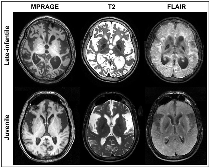
GM1-gangliosidosis (GM1) leads to extensive neurodegenerative changes and atrophy that precludes the use of automated MRI segmentation techniques for generating brain volumetrics. We developed a standardized segmentation protocol for brain MRIs of patients with type II GM1 and then assessed the inter- and intra-rater reliability of this methodology. The volumetric data may be used as a biomarker of disease burden and progression, and standardized methodology may support research into the natural history of the disease which is currently lacking in the literature.
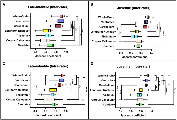
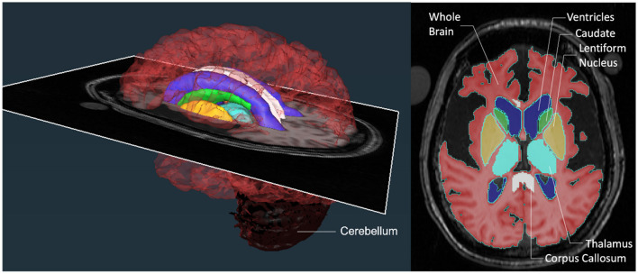
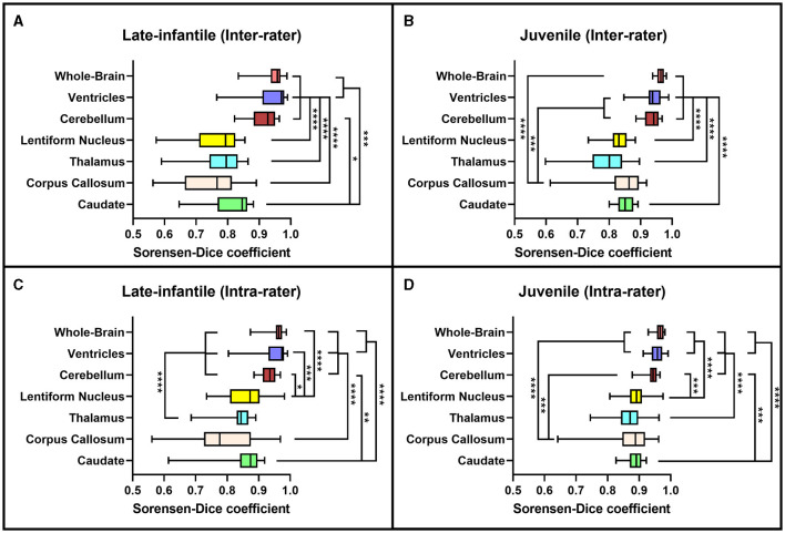
Twenty-five brain MRIs were included in this study from 22 type II GM1 patients of which 8 were late-infantile subtype and 14 were juvenile subtype. The following structures were segmented by two rating teams on a slice-by-slice basis: whole brain, ventricles, cerebellum, lentiform nucleus, thalamus, corpus callosum, and caudate nucleus. The inter- and intra-rater reliability of the segmentation method was assessed with an intraclass correlation coefficient as well as Sorensen-Dice and Jaccard coefficients.
Based on the Sorensen-Dice and Jaccard coefficients, the inter- and intra-rater reliability of the segmentation method was significantly better for the juvenile patients compared to late-infantile ( < 0.01). In addition, the agreement between the two rater teams and within themselves can be considered good with all -values < 0.05.
The standardized segmentation approach described here has good inter- and intra-rater reliability and may provide greater accuracy and reproducibility for neuromorphological studies in this group of patients and help to further expand our understanding of the natural history of this disease.
GM1神经节苷脂沉积症(GM1)会导致广泛的神经退行性改变和萎缩,这使得无法使用自动MRI分割技术来生成脑容量数据。我们为II型GM1患者的脑部MRI开发了一种标准化分割方案,然后评估了该方法在评分者间和评分者内的可靠性。容量数据可作为疾病负担和进展的生物标志物,标准化方法可能有助于对该疾病自然史的研究,而目前文献中缺乏这方面的研究。
本研究纳入了22例II型GM1患者的25份脑部MRI,其中8例为晚婴型亚型,14例为青少年型亚型。两个评分团队逐片分割以下结构:全脑、脑室、小脑、豆状核、丘脑、胼胝体和尾状核。使用组内相关系数以及索伦森-戴斯系数和杰卡德系数评估分割方法在评分者间和评分者内的可靠性。
基于索伦森-戴斯系数和杰卡德系数,与晚婴型患者相比,青少年患者分割方法在评分者间和评分者内的可靠性显著更高(<0.01)。此外,两个评分团队之间以及团队内部的一致性均可认为良好,所有p值<0.05。
本文所述的标准化分割方法在评分者间和评分者内具有良好的可靠性,可能为该组患者的神经形态学研究提供更高的准确性和可重复性,并有助于进一步扩展我们对该疾病自然史的理解。