• 文献检索
  • 文档翻译
  • 深度研究
  • 学术资讯
  • Suppr Zotero 插件Zotero 插件
  • 邀请有礼
  • 套餐&价格
  • 历史记录
应用&插件
Suppr Zotero 插件Zotero 插件浏览器插件Mac 客户端Windows 客户端微信小程序
定价
高级版会员购买积分包购买API积分包
服务
文献检索文档翻译深度研究API 文档MCP 服务
关于我们
关于 Suppr公司介绍联系我们用户协议隐私条款
关注我们

Suppr 超能文献

核心技术专利:CN118964589B侵权必究
粤ICP备2023148730 号-1Suppr @ 2026

文献检索

告别复杂PubMed语法,用中文像聊天一样搜索,搜遍4000万医学文献。AI智能推荐,让科研检索更轻松。

立即免费搜索

文件翻译

保留排版,准确专业,支持PDF/Word/PPT等文件格式,支持 12+语言互译。

免费翻译文档

深度研究

AI帮你快速写综述,25分钟生成高质量综述,智能提取关键信息,辅助科研写作。

立即免费体验

肠微绒毛细胞顶域细胞骨架超微结构的组织。

Organization of a cytoskeletal superstructure in the apical domain of intestinal tuft cells.

机构信息

Department of Cell and Developmental Biology, Vanderbilt University School of Medicine, Nashville, TN, USA.

Vanderbilt Cell Imaging Shared Resource, Vanderbilt University , Nashville, TN, USA.

出版信息

J Cell Biol. 2024 Dec 2;223(12). doi: 10.1083/jcb.202404070. Epub 2024 Oct 1.

DOI:10.1083/jcb.202404070
PMID:39352498
原文链接:https://pmc.ncbi.nlm.nih.gov/articles/PMC11457492/
Abstract

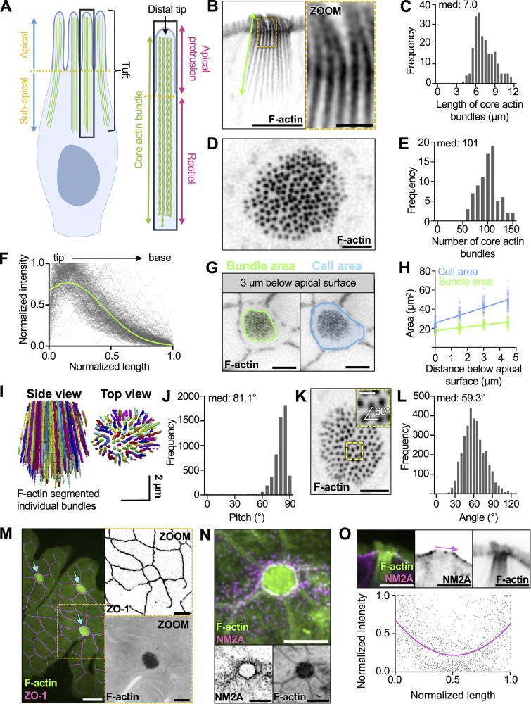
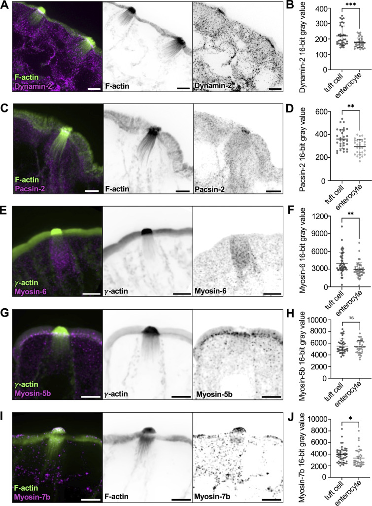
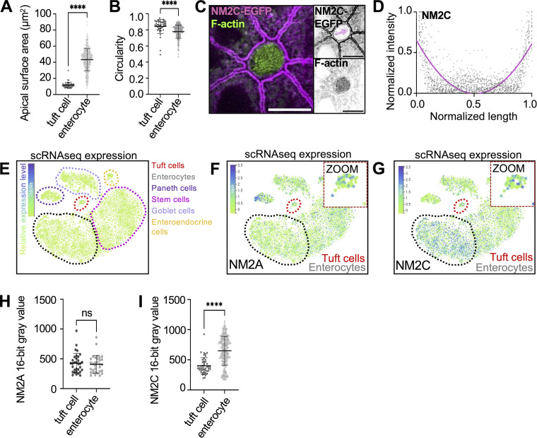
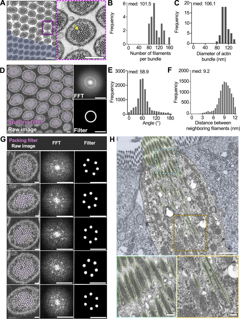
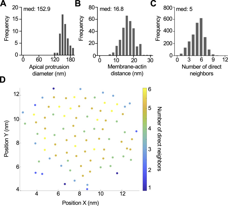
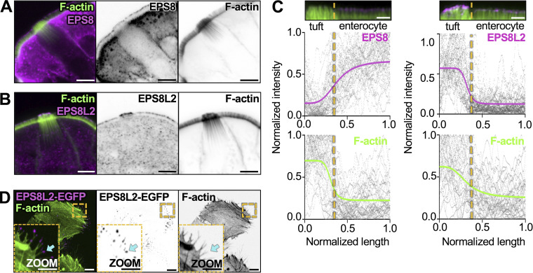
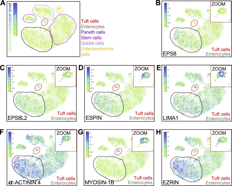
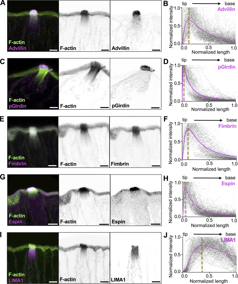
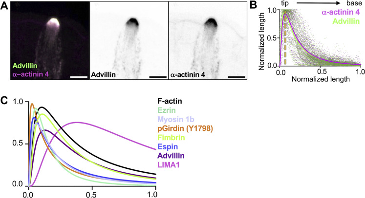
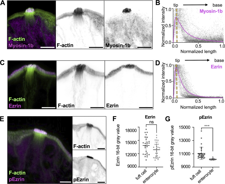
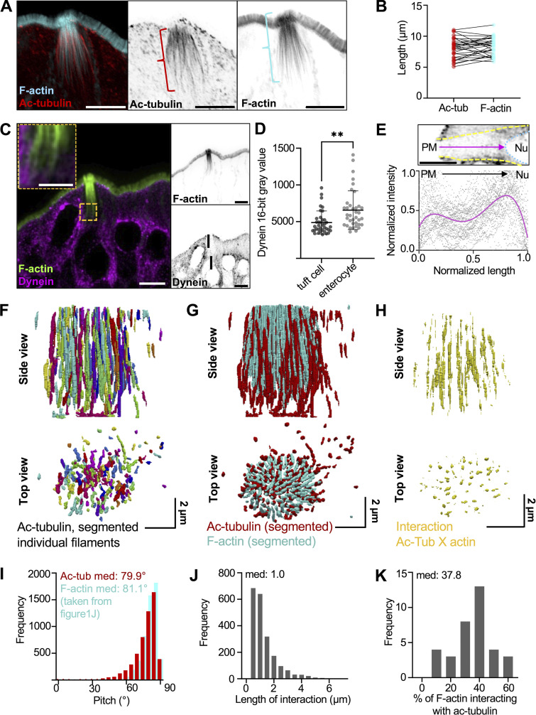
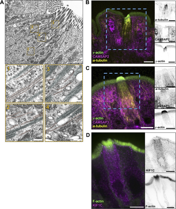
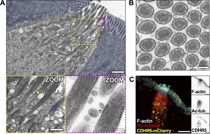
Tuft cells are a rare epithelial cell type that play important roles in sensing and responding to luminal antigens. A defining morphological feature of this lineage is the actin-rich apical "tuft," which contains large fingerlike protrusions. However, details of the cytoskeletal ultrastructure underpinning the tuft, the molecules involved in building this structure, or how it supports tuft cell biology remain unclear. In the context of the small intestine, we found that tuft cell protrusions are supported by long-core bundles that consist of F-actin crosslinked in a parallel and polarized configuration; they also contain a tuft cell-specific complement of actin-binding proteins that exhibit regionalized localization along the bundle axis. Remarkably, in the sub-apical cytoplasm, the array of core actin bundles interdigitates and co-aligns with a highly ordered network of microtubules. The resulting cytoskeletal superstructure is well positioned to support subcellular transport and, in turn, the dynamic sensing functions of the tuft cell that are critical for intestinal homeostasis.

摘要

类指突细胞是一种罕见的上皮细胞类型,在感知和响应腔抗原中发挥重要作用。该谱系的一个定义性形态特征是富含肌动蛋白的顶端“类指突”,其中包含大的指状突起。然而,支持类指突的细胞骨架超微结构的细节、参与构建该结构的分子,或它如何支持类指突细胞生物学仍然不清楚。在小肠的背景下,我们发现类指突细胞的突起由长核心束支撑,这些核心束由平行且极化排列的 F-actin 交联而成;它们还包含一组特定的肌动蛋白结合蛋白,这些蛋白在沿着束轴的区域化定位。值得注意的是,在亚顶细胞质中,核心肌动蛋白束的排列与高度有序的微管网络交织并对齐。由此产生的细胞骨架超结构非常适合支持细胞内运输,进而支持类指突细胞的动态感应功能,这对于肠道内稳态至关重要。

https://cdn.ncbi.nlm.nih.gov/pmc/blobs/ca70/11457492/ffd8feb3c0a1/JCB_202404070_Fig8.jpg
https://cdn.ncbi.nlm.nih.gov/pmc/blobs/ca70/11457492/576b2f407e30/JCB_202404070_Fig1.jpg
https://cdn.ncbi.nlm.nih.gov/pmc/blobs/ca70/11457492/76e7d3ffe597/JCB_202404070_FigS1.jpg
https://cdn.ncbi.nlm.nih.gov/pmc/blobs/ca70/11457492/3fe34a22aa98/JCB_202404070_Fig2.jpg
https://cdn.ncbi.nlm.nih.gov/pmc/blobs/ca70/11457492/3337260a1774/JCB_202404070_FigS2.jpg
https://cdn.ncbi.nlm.nih.gov/pmc/blobs/ca70/11457492/5e54ff231083/JCB_202404070_Fig3.jpg
https://cdn.ncbi.nlm.nih.gov/pmc/blobs/ca70/11457492/ff8a1ae34c43/JCB_202404070_FigS3.jpg
https://cdn.ncbi.nlm.nih.gov/pmc/blobs/ca70/11457492/677a758af93d/JCB_202404070_Fig4.jpg
https://cdn.ncbi.nlm.nih.gov/pmc/blobs/ca70/11457492/19c4259c1042/JCB_202404070_FigS4.jpg
https://cdn.ncbi.nlm.nih.gov/pmc/blobs/ca70/11457492/d779e4ce3eb3/JCB_202404070_Fig5.jpg
https://cdn.ncbi.nlm.nih.gov/pmc/blobs/ca70/11457492/8f9ec0af1028/JCB_202404070_Fig6.jpg
https://cdn.ncbi.nlm.nih.gov/pmc/blobs/ca70/11457492/898c8792b43d/JCB_202404070_FigS5.jpg
https://cdn.ncbi.nlm.nih.gov/pmc/blobs/ca70/11457492/4df64a8bffdd/JCB_202404070_Fig7.jpg
https://cdn.ncbi.nlm.nih.gov/pmc/blobs/ca70/11457492/ffd8feb3c0a1/JCB_202404070_Fig8.jpg

相似文献

1
Organization of a cytoskeletal superstructure in the apical domain of intestinal tuft cells.肠微绒毛细胞顶域细胞骨架超微结构的组织。
J Cell Biol. 2024 Dec 2;223(12). doi: 10.1083/jcb.202404070. Epub 2024 Oct 1.
2
Intestinal tuft cells assemble a cytoskeletal superstructure composed of co-aligned actin bundles and microtubules.肠道簇细胞组装由共排列的肌动蛋白束和微管组成的细胞骨架超结构。
bioRxiv. 2024 Mar 19:2024.03.19.585757. doi: 10.1101/2024.03.19.585757.
3
A CD317/tetherin-RICH2 complex plays a critical role in the organization of the subapical actin cytoskeleton in polarized epithelial cells.CD317/栓系蛋白-RICH2复合物在极化上皮细胞的顶端下肌动蛋白细胞骨架组织中起关键作用。
J Cell Biol. 2009 Mar 9;184(5):721-36. doi: 10.1083/jcb.200804154.
4
Sperm Flagellar 1 Binds Actin in Intestinal Epithelial Cells and Contributes to Formation of Filopodia and Lamellipodia.精子鞭毛蛋白 1 在肠道上皮细胞中结合肌动蛋白,并有助于形成丝状伪足和片状伪足。
Gastroenterology. 2019 Dec;157(6):1544-1555.e3. doi: 10.1053/j.gastro.2019.08.031. Epub 2019 Aug 29.
5
The cortical cytoskeletal network and cell-wall dynamics in the unicellular charophycean green alga Penium margaritaceum.单细胞轮藻纲绿藻Margaritaceum中皮层细胞骨架网络和细胞壁动态变化
Ann Bot. 2014 Oct;114(6):1237-49. doi: 10.1093/aob/mcu013. Epub 2014 Mar 5.
6
Actin-interacting protein 1 controls assembly and permeability of intestinal epithelial apical junctions.肌动蛋白相互作用蛋白1控制肠上皮顶端连接的组装和通透性。
Am J Physiol Gastrointest Liver Physiol. 2015 May 1;308(9):G745-56. doi: 10.1152/ajpgi.00446.2014. Epub 2015 Mar 19.
7
In vivo, villin is required for Ca(2+)-dependent F-actin disruption in intestinal brush borders.在体内,绒毛蛋白是小肠刷状缘中钙依赖型F-肌动蛋白破坏所必需的。
J Cell Biol. 1999 Aug 23;146(4):819-30. doi: 10.1083/jcb.146.4.819.
8
In vitro reconstitution of dynamic microtubules interacting with actin filament networks.与肌动蛋白丝网络相互作用的动态微管的体外重建。
Methods Enzymol. 2014;540:301-20. doi: 10.1016/B978-0-12-397924-7.00017-0.
9
Tropomyosin distinguishes between the two actin-binding sites of villin and affects actin-binding properties of other brush border proteins.原肌球蛋白可区分绒毛蛋白的两个肌动蛋白结合位点,并影响其他刷状缘蛋白的肌动蛋白结合特性。
J Cell Biol. 1987 Jan;104(1):29-40. doi: 10.1083/jcb.104.1.29.
10
Intestinal Tuft Cells: Morphology, Function, and Implications for Human Health.肠簇细胞:形态、功能及其对人类健康的影响。
Annu Rev Physiol. 2024 Feb 12;86:479-504. doi: 10.1146/annurev-physiol-042022-030310. Epub 2023 Oct 20.

本文引用的文献

1
ARHGAP18-ezrin functions as an autoregulatory module for RhoA in the assembly of distinct actin-based structures.ARHGAP18-埃兹蛋白通过形成不同的基于肌动蛋白的结构作为 RhoA 的自身调节模块发挥作用。
Elife. 2024 Jan 9;13:e83526. doi: 10.7554/eLife.83526.
2
The actin cytoskeleton in hair bundle development and hearing loss.毛细胞发育和听力损失中的肌动蛋白细胞骨架。
Hear Res. 2023 Sep 1;436:108817. doi: 10.1016/j.heares.2023.108817. Epub 2023 May 26.
3
Building the brush border, one microvillus at a time.一次一个微绒毛,构建刷状缘。
Curr Opin Cell Biol. 2023 Feb;80:102153. doi: 10.1016/j.ceb.2023.102153. Epub 2023 Feb 22.
4
Gut Epithelial Inositol Polyphosphate Multikinase Alleviates Experimental Colitis via Governing Tuft Cell Homeostasis.肠上皮肌醇多磷酸激酶通过调控微绒毛细胞稳态缓解实验性结肠炎。
Cell Mol Gastroenterol Hepatol. 2022;14(6):1235-1256. doi: 10.1016/j.jcmgh.2022.08.004. Epub 2022 Aug 19.
5
Force generation of KIF1C is impaired by pathogenic mutations.致病突变会损害 KIF1C 的产生力。
Curr Biol. 2022 Sep 12;32(17):3862-3870.e6. doi: 10.1016/j.cub.2022.07.029. Epub 2022 Aug 11.
6
The membrane-actin linker ezrin acts as a sliding anchor.膜 - 肌动蛋白连接蛋白埃兹蛋白作为滑动锚起作用。
Sci Adv. 2022 Aug 5;8(31):eabo2779. doi: 10.1126/sciadv.abo2779.
7
Dissociation and inDrops microfluidic encapsulation of human gut tissues for single-cell atlasing studies.人肠道组织的解离和 inDrop 微流控包封用于单细胞图谱研究。
STAR Protoc. 2022 Jul 19;3(3):101570. doi: 10.1016/j.xpro.2022.101570. eCollection 2022 Sep 16.
8
Unique and redundant functions of cytoplasmic actins and nonmuscle myosin II isoforms at epithelial junctions.细胞质肌动蛋白和非肌肉肌球蛋白 II 同工型在细胞连接中的独特和冗余功能。
Ann N Y Acad Sci. 2022 Sep;1515(1):61-74. doi: 10.1111/nyas.14808. Epub 2022 Jun 7.
9
Mitotic Spindle Positioning (MISP) is an actin bundler that selectively stabilizes the rootlets of epithelial microvilli.有丝分裂纺锤体定位(MISP)是一种肌动蛋白束形成蛋白,它选择性地稳定上皮微绒毛的根丝。
Cell Rep. 2022 Apr 19;39(3):110692. doi: 10.1016/j.celrep.2022.110692.
10
PGD2 and CRTH2 counteract Type 2 cytokine-elicited intestinal epithelial responses during helminth infection.PGD2 和 CRTH2 可拮抗寄生虫感染期间 2 型细胞因子诱导的肠道上皮细胞反应。
J Exp Med. 2021 Sep 6;218(9). doi: 10.1084/jem.20202178. Epub 2021 Jul 20.