• 文献检索
  • 文档翻译
  • 深度研究
  • 学术资讯
  • Suppr Zotero 插件Zotero 插件
  • 邀请有礼
  • 套餐&价格
  • 历史记录
应用&插件
Suppr Zotero 插件Zotero 插件浏览器插件Mac 客户端Windows 客户端微信小程序
定价
高级版会员购买积分包购买API积分包
服务
文献检索文档翻译深度研究API 文档MCP 服务
关于我们
关于 Suppr公司介绍联系我们用户协议隐私条款
关注我们

Suppr 超能文献

核心技术专利:CN118964589B侵权必究
粤ICP备2023148730 号-1Suppr @ 2026

文献检索

告别复杂PubMed语法,用中文像聊天一样搜索,搜遍4000万医学文献。AI智能推荐,让科研检索更轻松。

立即免费搜索

文件翻译

保留排版,准确专业,支持PDF/Word/PPT等文件格式,支持 12+语言互译。

免费翻译文档

深度研究

AI帮你快速写综述,25分钟生成高质量综述,智能提取关键信息,辅助科研写作。

立即免费体验

超声评估马掌骨软骨厚度比计算机断层扫描关节造影更准确。

Ultrasonographic assessment of equine metacarpal cartilage thickness is more accurate than computed tomographic arthrography.

作者信息

Hoey Seamus, Fogarty Ursula, McAllister Hester, Puggioni Antonella, Cloak Brian, Richard Hélène, Skelly Cliona, Laverty Sheila

机构信息

Equine Clinical Studies, Diagnostic Imaging and Anaesthesia, School of Veterinary Medicine, University College, Dublin, Dublin, Ireland.

Irish Equine Centre, Johnstown, Naas, Kildare, Ireland.

出版信息

Vet Radiol Ultrasound. 2025 Jan;66(1):e13444. doi: 10.1111/vru.13444. Epub 2024 Oct 4.

DOI:10.1111/vru.13444
PMID:39367616
原文链接:https://pmc.ncbi.nlm.nih.gov/articles/PMC11617610/
Abstract

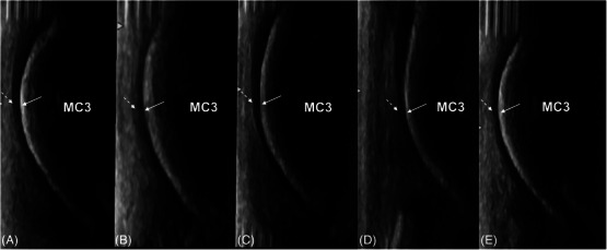
Articular cartilage can be directly imaged using ultrasonography. The fetlock is a common site of osteochondrosis, with the sagittal ridge of the third metacarpal bone most commonly affected. In osteochondrosis, cartilage thickening may be an initial finding. This postmortem study investigated the ability of ultrasonography to accurately measure the dorsodistal articular cartilage of the third metacarpal bone in young horses, compared to computed tomographic arthrography (CTA) and histological measurements. A total of 33 metacarpophalangeal joints from 18 horses between the ages of 12 days and 10 months old were imaged ultrasonographically and with CTA and sectioned and measured using histology. Imaging measurements were made by two observers. Despite overall weak agreement between ultrasonography and histology, the best agreement was at the distal aspect of the sagittal ridge of the third metacarpal bone. Interobserver agreement at this site was also moderate. CTA showed poor agreement overall with histology. Cartilage thickness decreased with age on ultrasonography, CTA, and histology. In conclusion, ultrasonography is a more accurate imaging modality than CTA in the assessment of cartilage in young horses.

摘要

关节软骨可用超声直接成像。跗关节是骨软骨病的常见部位,第三掌骨矢状嵴最常受累。在骨软骨病中,软骨增厚可能是最初的表现。这项尸检研究调查了与计算机断层扫描关节造影(CTA)和组织学测量相比,超声准确测量幼马第三掌骨背侧远端关节软骨的能力。对18匹年龄在12天至10个月之间的马的33个掌指关节进行了超声成像、CTA成像,并进行切片和组织学测量。成像测量由两名观察者进行。尽管超声与组织学之间总体一致性较弱,但最佳一致性出现在第三掌骨矢状嵴的远端。该部位观察者间的一致性也为中等。CTA总体上与组织学的一致性较差。超声、CTA和组织学检查均显示软骨厚度随年龄增长而降低。总之,在评估幼马软骨方面,超声是比CTA更准确的成像方式。

https://cdn.ncbi.nlm.nih.gov/pmc/blobs/62ff/11617610/3d0f2034d767/VRU-66-0-g008.jpg
https://cdn.ncbi.nlm.nih.gov/pmc/blobs/62ff/11617610/1e321ee9af4f/VRU-66-0-g004.jpg
https://cdn.ncbi.nlm.nih.gov/pmc/blobs/62ff/11617610/ee93fe83f2f3/VRU-66-0-g005.jpg
https://cdn.ncbi.nlm.nih.gov/pmc/blobs/62ff/11617610/465c74eb2435/VRU-66-0-g007.jpg
https://cdn.ncbi.nlm.nih.gov/pmc/blobs/62ff/11617610/a92575f8fdc7/VRU-66-0-g001.jpg
https://cdn.ncbi.nlm.nih.gov/pmc/blobs/62ff/11617610/49597b43e57d/VRU-66-0-g002.jpg
https://cdn.ncbi.nlm.nih.gov/pmc/blobs/62ff/11617610/f64587d51eb9/VRU-66-0-g006.jpg
https://cdn.ncbi.nlm.nih.gov/pmc/blobs/62ff/11617610/8e7329ee9002/VRU-66-0-g003.jpg
https://cdn.ncbi.nlm.nih.gov/pmc/blobs/62ff/11617610/3d0f2034d767/VRU-66-0-g008.jpg

相似文献

1
Ultrasonographic assessment of equine metacarpal cartilage thickness is more accurate than computed tomographic arthrography.超声评估马掌骨软骨厚度比计算机断层扫描关节造影更准确。
Vet Radiol Ultrasound. 2025 Jan;66(1):e13444. doi: 10.1111/vru.13444. Epub 2024 Oct 4.
2
CORRELATION OF ARTICULAR CARTILAGE THICKNESS MEASUREMENTS MADE WITH MAGNETIC RESONANCE IMAGING, MAGNETIC RESONANCE ARTHROGRAPHY, AND COMPUTED TOMOGRAPHIC ARTHROGRAPHY WITH GROSS ARTICULAR CARTILAGE THICKNESS IN THE EQUINE METACARPOPHALANGEAL JOINT.马掌指关节中磁共振成像、磁共振关节造影、计算机断层扫描关节造影所测关节软骨厚度与大体关节软骨厚度的相关性
Vet Radiol Ultrasound. 2016 Sep;57(5):515-25. doi: 10.1111/vru.12390. Epub 2016 Aug 1.
3
Comparison between magnetic resonance imaging, computed tomography, and arthrography to identify artificially induced cartilage defects of the equine carpal joints.磁共振成像、计算机断层扫描和关节造影术在识别马腕关节人工诱导软骨缺损方面的比较。
Vet Radiol Ultrasound. 2018 May;59(3):312-325. doi: 10.1111/vru.12598. Epub 2018 Feb 18.
4
Detection of articular pathology of the distal aspect of the third metacarpal bone in thoroughbred racehorses: comparison of radiography, computed tomography and magnetic resonance imaging.纯种赛马第三掌骨远端关节病变的检测:X线摄影、计算机断层扫描和磁共振成像的比较
Vet Surg. 2011 Dec;40(8):942-51. doi: 10.1111/j.1532-950X.2011.00881.x. Epub 2011 Oct 31.
5
A comparison of 3-T magnetic resonance imaging and computed tomography arthrography to identify structural cartilage defects of the fetlock joint in the horse.3-T磁共振成像与计算机断层扫描关节造影术在识别马跗关节结构软骨缺损中的比较。
Vet J. 2014 Jan;199(1):115-22. doi: 10.1016/j.tvjl.2013.10.021. Epub 2013 Oct 26.
6
Fat-suppressed spoiled gradient-recalled imaging of equine metacarpophalangeal articular cartilage.马掌指关节软骨的脂肪抑制扰相梯度回波成像
Vet Radiol Ultrasound. 2010 Mar-Apr;51(2):107-15. doi: 10.1111/j.1740-8261.2009.01633.x.
7
Cartilage defect identification on computed tomography arthrography in equine fetlock: Ex-vivo study.马球节理计算机断层扫描关节造影术中软骨缺损的识别:离体研究。
Res Vet Sci. 2025 Mar;185:105548. doi: 10.1016/j.rvsc.2025.105548. Epub 2025 Jan 21.
8
COMPARISON BETWEEN COMPUTED TOMOGRAPHIC ARTHROGRAPHY, RADIOGRAPHY, ULTRASONOGRAPHY, AND ARTHROSCOPY FOR THE DIAGNOSIS OF FEMOROTIBIAL JOINT DISEASE IN WESTERN PERFORMANCE HORSES.计算机断层扫描关节造影、X线摄影、超声检查和关节镜检查在西部表演马股胫关节疾病诊断中的比较
Vet Radiol Ultrasound. 2016 Jul;57(4):387-402. doi: 10.1111/vru.12366. Epub 2016 May 12.
9
A threshold volume of 10 ml is suggested for detecting articular cartilage defects in equine carpal joints using CT arthrography: Ex vivo pilot study.建议使用 CT 关节造影术检测马腕关节关节软骨缺损的阈值体积为 10ml:离体初步研究。
Vet Radiol Ultrasound. 2022 Jan;63(1):54-63. doi: 10.1111/vru.13028. Epub 2021 Oct 20.
10
Comparison of gross and histopathologic findings with quantitative computed tomographic bone density in the distal third metacarpal bone of racehorses.赛马第三掌骨远端大体及组织病理学检查结果与定量计算机断层扫描骨密度的比较。
Vet Radiol Ultrasound. 2007 Nov-Dec;48(6):518-27. doi: 10.1111/j.1740-8261.2007.00289.x.

本文引用的文献

1
Maturation of the equine medial femoral condyle osteochondral unit.马内侧股骨髁骨软骨单元的成熟
Osteoarthr Cartil Open. 2020 Jan 27;2(1):100029. doi: 10.1016/j.ocarto.2020.100029. eCollection 2020 Mar.
2
Ultrasound screening protocol for osteochondrosis at selected predilection sites in thoroughbred yearlings.纯种一岁马特定好发部位骨软骨病的超声筛查方案
Ir Vet J. 2022 Apr 27;75(1):8. doi: 10.1186/s13620-022-00216-7.
3
Imaging and Gross Pathological Appearance of Changes in the Parasagittal Grooves of Thoroughbred Racehorses.
纯种赛马矢状旁沟变化的影像学及大体病理学表现
Animals (Basel). 2021 Nov 24;11(12):3366. doi: 10.3390/ani11123366.
4
Septic Arthritis/Osteomyelitis May Lead to Osteochondrosis-Like Lesions in Foals.脓毒性关节炎/骨髓炎可能导致幼驹出现类似骨软骨病的病变。
Vet Pathol. 2018 Sep;55(5):693-702. doi: 10.1177/0300985818777786. Epub 2018 May 28.
5
Comparison between magnetic resonance imaging, computed tomography, and arthrography to identify artificially induced cartilage defects of the equine carpal joints.磁共振成像、计算机断层扫描和关节造影术在识别马腕关节人工诱导软骨缺损方面的比较。
Vet Radiol Ultrasound. 2018 May;59(3):312-325. doi: 10.1111/vru.12598. Epub 2018 Feb 18.
6
Ultrasonographic screening for subclinical osteochondrosis of the femoral trochlea in foals (28-166 days old): a prospective farm study.超声检查对驹(28 - 166日龄)股骨滑车亚临床骨软骨病的筛查:一项前瞻性农场研究
Equine Vet J. 2018 May;50(3):312-320. doi: 10.1111/evj.12772. Epub 2017 Nov 9.
7
Validation of the ultrasonographic assessment of the femoral trochlea epiphyseal cartilage in foals at osteochondrosis predilected sites with magnetic resonance imaging and histology.通过磁共振成像和组织学对马驹骨软骨病好发部位股骨滑车骨骺软骨超声评估的验证。
Equine Vet J. 2017 Nov;49(6):821-828. doi: 10.1111/evj.12698. Epub 2017 Jun 19.
8
CORRELATION OF ARTICULAR CARTILAGE THICKNESS MEASUREMENTS MADE WITH MAGNETIC RESONANCE IMAGING, MAGNETIC RESONANCE ARTHROGRAPHY, AND COMPUTED TOMOGRAPHIC ARTHROGRAPHY WITH GROSS ARTICULAR CARTILAGE THICKNESS IN THE EQUINE METACARPOPHALANGEAL JOINT.马掌指关节中磁共振成像、磁共振关节造影、计算机断层扫描关节造影所测关节软骨厚度与大体关节软骨厚度的相关性
Vet Radiol Ultrasound. 2016 Sep;57(5):515-25. doi: 10.1111/vru.12390. Epub 2016 Aug 1.
9
Heritability and prevalence of selected osteochondrosis lesions in yearling Thoroughbred horses.一岁纯种马特定骨软骨病病变的遗传力和患病率
Equine Vet J. 2017 May;49(3):282-287. doi: 10.1111/evj.12613. Epub 2016 Sep 4.
10
COMPARISON BETWEEN COMPUTED TOMOGRAPHIC ARTHROGRAPHY, RADIOGRAPHY, ULTRASONOGRAPHY, AND ARTHROSCOPY FOR THE DIAGNOSIS OF FEMOROTIBIAL JOINT DISEASE IN WESTERN PERFORMANCE HORSES.计算机断层扫描关节造影、X线摄影、超声检查和关节镜检查在西部表演马股胫关节疾病诊断中的比较
Vet Radiol Ultrasound. 2016 Jul;57(4):387-402. doi: 10.1111/vru.12366. Epub 2016 May 12.