Fong Henrick Ryan C, Zilbermint Mihail
Internal Medicine, Ascension Saint Agnes Hospital, Baltimore, USA.
Endocrinology, Diabetes and Metabolism, Suburban Hospital, Bethesda, USA.
Cureus. 2024 Sep 4;16(9):e68637. doi: 10.7759/cureus.68637. eCollection 2024 Sep.
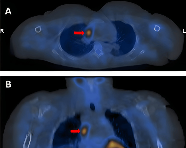
Ectopic parathyroid adenomas pose significant diagnostic and therapeutic challenges due to their atypical locations outside the usual anatomical boundaries of the parathyroid glands. These adenomas, which represent a small percentage of primary hyperparathyroidism cases, are often found in areas such as the mediastinum, thymus, or retroesophageal space. Their ectopic nature complicates diagnosis, as traditional neck imaging techniques may fail to localize these glands. We present the case of a 27-year-old female who initially presented with nausea, vomiting, severe hypercalcemia, and elevated parathyroid hormone (PTH) levels. Despite being advised to consult an endocrinologist, she experienced difficulty scheduling an appointment. Due to persistent symptoms and laboratory abnormalities, she was subsequently admitted to the hospital. Initial neck imaging failed to identify the parathyroid adenoma. However, subsequent imaging, including parathyroid scintigraphy, revealed an ectopic parathyroid adenoma located in the mediastinum. The patient underwent a successful robotically assisted thymectomy, guided by intraoperative PTH monitoring, which resulted in the resolution of hypercalcemia and normalization of PTH levels. This case underscores the importance of a comprehensive diagnostic approach when dealing with ectopic parathyroid adenomas. Parathyroid scintigraphy, in particular, proves to be a critical tool due to its high sensitivity in detecting ectopic glands. Moreover, our findings emphasize the need for a high index of suspicion for ectopic parathyroid adenomas, especially when conventional neck imaging is inconclusive in cases of hyperparathyroidism. Timely and accurate diagnosis is essential for facilitating precise surgical intervention, ultimately leading to improved patient outcomes.
异位甲状旁腺腺瘤因其位于甲状旁腺正常解剖边界之外的非典型位置,带来了重大的诊断和治疗挑战。这些腺瘤占原发性甲状旁腺功能亢进病例的比例较小,常发现于纵隔、胸腺或食管后间隙等部位。它们的异位性质使诊断变得复杂,因为传统的颈部成像技术可能无法定位这些腺体。我们报告一例27岁女性病例,她最初表现为恶心、呕吐、严重高钙血症和甲状旁腺激素(PTH)水平升高。尽管有人建议她咨询内分泌科医生,但她在安排预约时遇到困难。由于症状持续和实验室检查异常,她随后入院。最初的颈部成像未能识别出甲状旁腺腺瘤。然而,随后的成像检查,包括甲状旁腺闪烁显像,显示在纵隔中有一个异位甲状旁腺腺瘤。患者在术中PTH监测的引导下,成功接受了机器人辅助胸腺切除术,高钙血症得以缓解,PTH水平恢复正常。该病例强调了处理异位甲状旁腺腺瘤时采用全面诊断方法的重要性。特别是甲状旁腺闪烁显像,因其在检测异位腺体方面具有高敏感性,被证明是一种关键工具。此外,我们的研究结果强调,对于异位甲状旁腺腺瘤需要高度怀疑,尤其是在甲状旁腺功能亢进病例中传统颈部成像结果不明确时。及时准确的诊断对于促进精确的手术干预至关重要,最终可改善患者预后。