Jung Dae Jin, Kim Gwang Ha, Kim Kyungbin, Jeon Hye Kyung, Joo Dong Chan, Lee Moon Won, Lee Bong Eun
Department of Internal Medicine, Pusan National University School of Medicine and Biomedical Research Institute, Pusan National University Hospital, Busan, Korea.
Department of Pathology, Pusan National University Hospital, Busan, Korea.
Gut Liver. 2025 Jan 15;19(1):50-58. doi: 10.5009/gnl240097. Epub 2024 Oct 8.
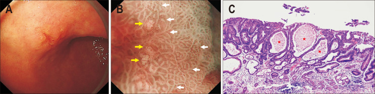
BACKGROUND/AIMS: Magnifying endoscopy with narrow-band imaging (ME-NBI) enables the visualization of detailed microsurface (MS) and microvascular (MV) structures in the gastrointestinal tract. White globe appearance (WGA) is a small whitish lesion with a globular shape identified during ME-NBI for early gastric cancer (EGC). This study aimed to investigate the associations between WGA, clinicopathological characteristics, and other ME-NBI findings in patients with EGC.
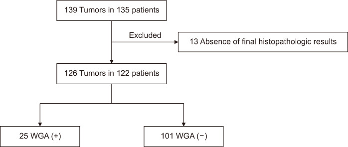
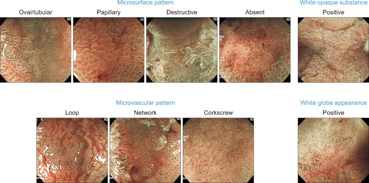
The presence or absence of WGA in 122 patients (126 lesions) with an endoscopic diagnosis of EGC who underwent ME-NBI before endoscopic or surgical resection was prospectively collected and retrospectively analyzed. During ME-NBI, the MS and MV patterns and the presence of WGA and white opaque substances (WOS) were investigated. EGC cases were categorized as differentiated or undifferentiated type, and mucosal, submucosal, or advanced.
Of 126 lesions, WGA was observed in 25 (19.8%). WGA was associated with tumor size (≤2 cm [17/63, 27.0%] vs >2 cm [8/63, 12.7%]; p=0.044), histologic type (differentiated type [22/89, 24.7%] vs undifferentiated type [3/37. 8.1%]; p=0.033), and tumor location (upper third [1/11, 9.1%] vs middle third [18/58, 31.0%] and lower third [6/57, 10.5%]; p=0.017). Although WGA was observed more frequently in lesions with an oval/tubular MS pattern, a fine-network MV pattern, and the absence of WOS, the difference was not statistically significant (MS pattern, p=0.358; MV pattern, p=0.212; WOS, p=0.121, respectively).
WGA was associated with small tumor size, differentiated-type histology, and middle-third tumor location, and was more frequently observed in lesions with an oval/tubular MS and fine-network MV patterns and the absence of WOS.
背景/目的:窄带成像放大内镜检查(ME-NBI)能够显示胃肠道详细的微表面(MS)和微血管(MV)结构。白色球状物外观(WGA)是在ME-NBI检查早期胃癌(EGC)时发现的一种球状小白色病变。本研究旨在探讨EGC患者中WGA、临床病理特征及其他ME-NBI表现之间的关联。
前瞻性收集并回顾性分析122例(126个病变)经内镜诊断为EGC且在接受内镜或手术切除前进行了ME-NBI检查的患者中WGA的有无情况。在ME-NBI检查过程中,观察MS和MV形态以及WGA和白色不透明物质(WOS)的存在情况。EGC病例分为分化型或未分化型,以及黏膜层、黏膜下层或进展期。
126个病变中,25个(19.8%)观察到WGA。WGA与肿瘤大小(≤2 cm [17/63, 27.0%] 对比>2 cm [8/63, 12.7%];p = 0.044)、组织学类型(分化型 [22/89, 24.7%] 对比未分化型 [3/37, 8.1%];p = 0.033)和肿瘤位置(上三分之一 [1/11, 9.1%] 对比中三分之一 [18/58, 31.0%] 和下三分之一 [6/57, 10.5%];p = 0.017)相关。尽管在具有椭圆形/管状MS形态、细网状MV形态且无WOS的病变中WGA更常被观察到,但差异无统计学意义(MS形态,p = 0.358;MV形态,p = 0.212;WOS,p = 0.121)。
WGA与肿瘤体积小、分化型组织学及中三分之一肿瘤位置相关,且在具有椭圆形/管状MS和细网状MV形态且无WOS的病变中更常被观察到。