Harvard Medical School, Boston, MA, 02115, USA.
Center for Advanced Orthopedic Studies, Beth Israel Deaconess Medical Center, Boston, MA, 02215, USA.
Sci Rep. 2024 Oct 8;14(1):23379. doi: 10.1038/s41598-024-75363-8.
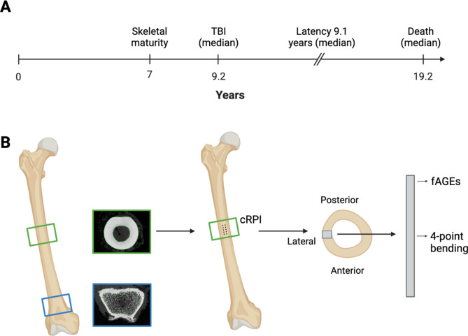
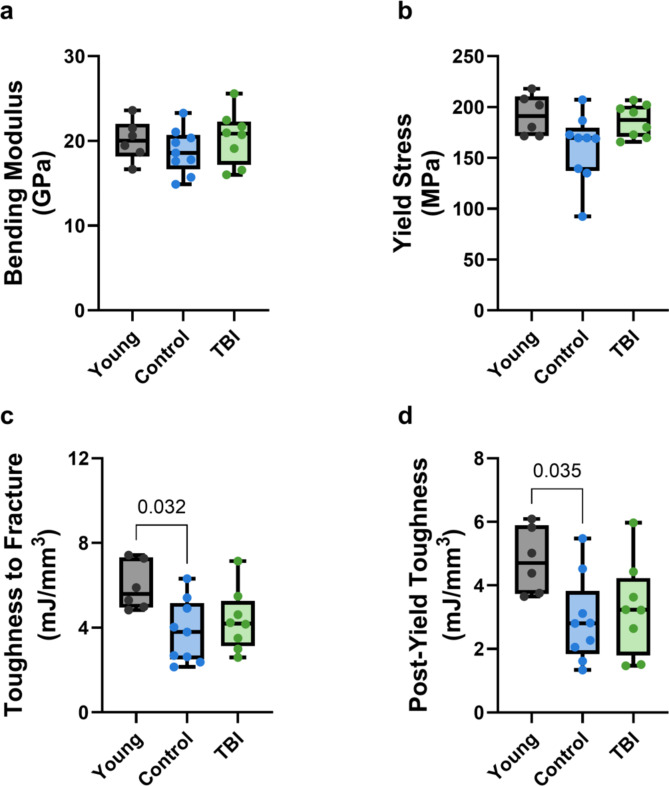
Exposure to ionizing radiation for oncological therapy increases the risk for late-onset fractures in survivors. However, the effects of total body irradiation (TBI) on adult bone are not well-characterized. The primary aim of this study was to quantify the long-term effects of TBI on bone microstructure, material composition, and mechanical behavior in skeletally mature rhesus macaque (Macaca mulatta) non-human primates. Femora were obtained post-mortem from animals exposed to an acute dose of TBI (6.0-6.75 Gy) nearly a decade earlier, age-matched non-irradiated controls, and non-irradiated young animals. The microstructure of femoral trabecular and cortical bone was assessed via micro-computed tomography. Material composition was evaluated by measuring total fluorescent advanced glycation end products (fAGEs). Cortical bone mechanical behavior was quantified via four-point bending and cyclic reference point indentation (cRPI). Animals exposed to TBI had slightly worse cortical microstructure, including lower cortical thickness (-11%, p = 0.037) and cortical area (-24%, p = 0.049), but similar fAGE content and mechanical properties as age-matched controls. Aging did not influence cortical microstructure, fAGE content, or cRPI measures but diminished femoral cortical post-yield properties, including toughness to fracture (-32%, p = 0.032). Because TBI was administered after the acquisition of peak bone mass, these results suggest that the skeletons of long-term survivors of adulthood TBI may be resilient, retaining or recovering their mechanical integrity during the post-treatment period, despite radiation-induced architectural deficits. Further investigation is necessary to better understand radiation-induced skeletal fragility in mature and immature bone to improve care for radiation patients of all ages.
接受用于肿瘤治疗的电离辐射会增加幸存者晚期骨折的风险。然而,全身照射(TBI)对成人骨骼的影响尚未得到很好的描述。本研究的主要目的是定量研究 TBI 对骨骼成熟恒河猴(Macaca mulatta)非人类灵长类动物的骨小梁和皮质骨微观结构、材料组成和力学性能的长期影响。在近十年前接受过急性 TBI(6.0-6.75 Gy)照射的动物死后获得股骨,与年龄匹配的未照射对照动物和未照射年轻动物一起进行研究。通过微计算机断层扫描评估股骨小梁和皮质骨的微观结构。通过测量总荧光晚期糖基化终产物(fAGE)来评估材料组成。通过四点弯曲和循环参考点压痕(cRPI)来量化皮质骨力学性能。接受 TBI 照射的动物皮质骨微观结构稍差,包括皮质骨厚度降低(-11%,p=0.037)和皮质骨面积减少(-24%,p=0.049),但 fAGE 含量和机械性能与年龄匹配的对照组相似。衰老不会影响皮质骨微观结构、fAGE 含量或 cRPI 测量值,但会降低股骨皮质的屈服后特性,包括抗断裂韧性(-32%,p=0.032)。由于 TBI 是在获得峰值骨量后给予的,因此这些结果表明,成年 TBI 长期幸存者的骨骼可能具有弹性,在治疗后期间保留或恢复其机械完整性,尽管存在辐射诱导的结构缺陷。需要进一步研究以更好地了解成熟和未成熟骨骼中的辐射诱导骨骼脆弱性,从而改善所有年龄段的辐射患者的护理。