Centre Européen du Cheval, Mont-le-Soie, Yvré-l'Évêque, Vielsalm.
Département des Sciences Cliniques des Équidés, Chirurgie et Orthopédie, FARAH, Université de Liège, Liège, Belgium.
PLoS One. 2024 Oct 11;19(10):e0311965. doi: 10.1371/journal.pone.0311965. eCollection 2024.
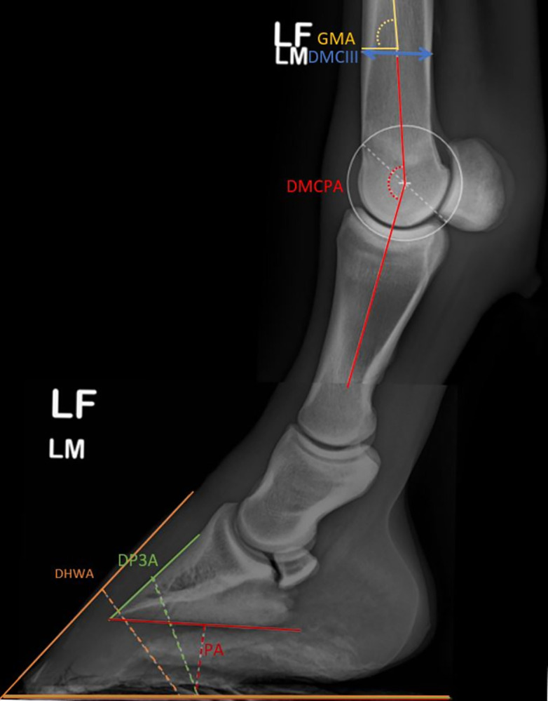
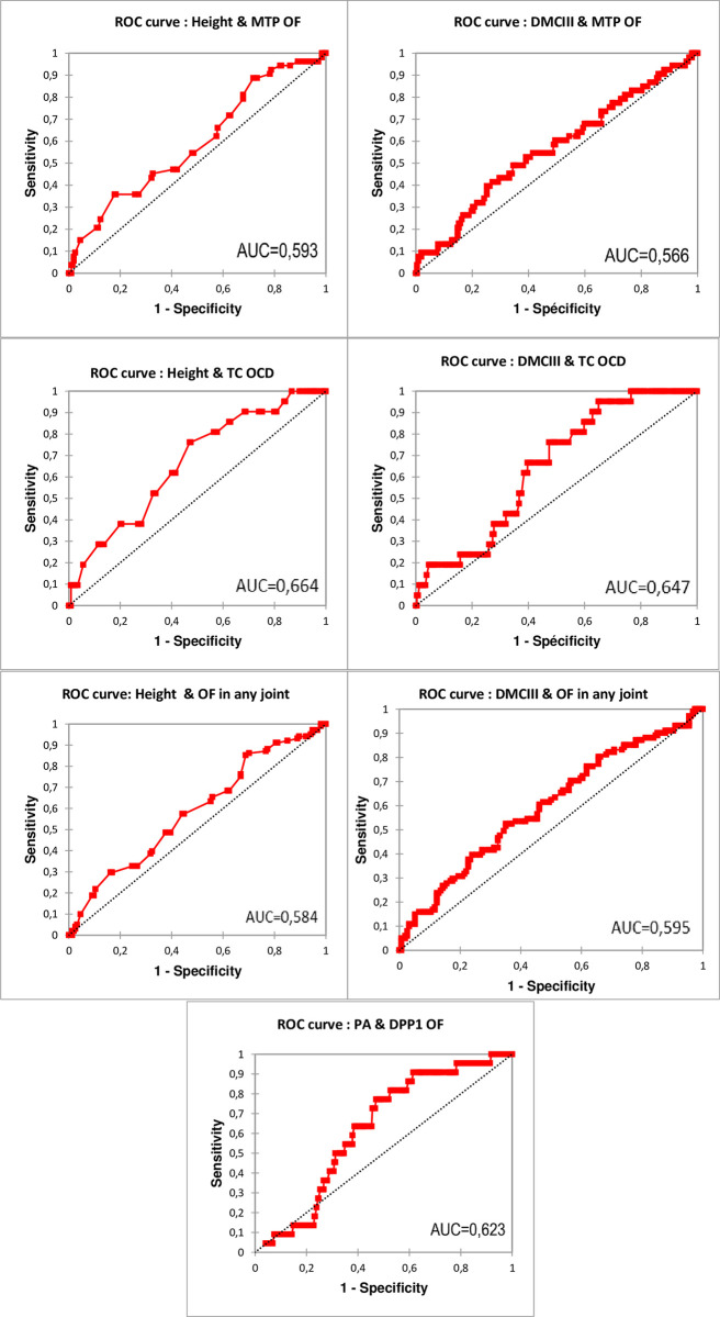
Osteochondral fragments within equine joints are commonly encountered and may predispose to lameness and limitation to sport purposes. Factors leading to this condition include genetic, nutritional and environmental conditions. However, few studies have evaluated the impact of conformation traits and their correlation with osteochondrosis. This study, based on the radiographic screenings of young horses born in Wallonia (266 individuals, 532 forelimbs), evaluated the correlation between foot, fetlock conformations of the front limb, height at the withers and presence of osteochondral fragments. Moreover, for all traits significantly associated with the presence of osteochondral fragments, a Receiver Operator Characteristic (ROC) curve, area under the curve and optimal cut-off value were calculated to predict the occurrence of fragments. Mean dorsal hoof wall angle was 52.36°, dorsal and palmar angle of the third phalanx were respectively 49.83° and 2.99°, and dorsal metacarpophalangeal angle 147.99°. Moreover, the prevalence of upright feet, defined as having an inclined profile of >2° steeper in relation to its contralateral counterpart, was 24%. Increased palmar angle of the distal phalanx was significantly correlated (P < 0.05) with presence of fragments located at the dorso-proximal margin of the proximal phalanx. The associated area under the curve was 0.623 (95% CI: 0528-0.717, P < 0.05) and the optimal cut-off value to predict fragment occurrence was 2.95° (sensitivity 77.3%; specificity 52.9%). Furthermore, the third metacarpal bone diameter of the left forelimb and height at the withers were significantly (P < 0.05) correlated with the presence of osteochondral fragments in general and within tarsocrural and metatarsophalangeal joints specifically. The area under the curve was 0.585 (95% CI: 0.513-0.656, P < 0.05) with an optimal cut-off value of 152.5 cm (sensitivity 85.1%; specificity 31.2%) for height at the withers to predict presence of osteochondral fragment; to predict the occurrence of osteochondral fragment in any joint on the basis of the third metacarpal bone diameter, the area under the curve was 0.595 (95% CI: 0.524-0.667, P <0.05) and the optimal cut-off value 34.9 mm (sensitivity 52.5%; specificity 64.9%). This study provides information about phenotypic traits associated with osteochondral fragments in horses. Although the diagnostic accuracy of these traits to detect osteochondral fragment was limited, the identification of more phenotypic characteristics could, in the future, make it possible to generate models for accurately identifying individuals at high risk of osteochondral fragments on the basis of their phenotype.
马关节内的软骨骨片很常见,可能导致跛行和运动受限。导致这种情况的因素包括遗传、营养和环境条件。然而,很少有研究评估形态特征及其与骨软骨病的相关性。本研究基于瓦隆(266 匹马,532 前肢)出生的年轻马的放射筛检,评估了前肢的足部、球节、肩高和软骨骨片存在之间的相关性。此外,对于与软骨骨片存在显著相关的所有特征,计算了接收者操作特征(ROC)曲线、曲线下面积和最佳截断值,以预测骨片的发生。背侧蹄壁角度平均值为 52.36°,第三跖骨的背侧和掌侧角度分别为 49.83°和 2.99°,掌侧掌指关节角度为 147.99°。此外,定义为相对于对侧有 2°以上倾斜度的直立足的比例为 24%。远端跖骨掌侧角度增加与近端骨片背侧-近端缘的骨片存在显著相关(P<0.05)。相关的曲线下面积为 0.623(95%CI:0.528-0.717,P<0.05),预测骨片发生的最佳截断值为 2.95°(灵敏度 77.3%;特异性 52.9%)。此外,左侧前肢的第三掌骨直径和肩高与跗跖关节和跖间关节中软骨骨片的存在显著相关(P<0.05)。曲线下面积为 0.585(95%CI:0.513-0.656,P<0.05),最佳截断值为 152.5cm(灵敏度 85.1%;特异性 31.2%),用于预测肩高是否存在软骨骨片;基于第三掌骨直径预测任何关节的软骨骨片发生,曲线下面积为 0.595(95%CI:0.524-0.667,P<0.05),最佳截断值为 34.9mm(灵敏度 52.5%;特异性 64.9%)。本研究提供了与马软骨骨片相关的表型特征信息。尽管这些特征在检测软骨骨片方面的诊断准确性有限,但未来识别更多的表型特征可能使基于表型识别高风险软骨骨片个体的模型成为可能。