Nozdriukhin Daniil, Cattaneo Marco, Klingler Norman, Lyu Shuxin, Li Weiye, de Espinosa Francisco Montero, Bonvin Jerome, Supponen Outi, Razansky Daniel, Deán-Ben Xosé Luís
Institute for Biomedical Engineering and Institute of Pharmacology and Toxicology, Faculty of Medicine, University of Zürich, Winterthurerstrasse 190, Zurich, 8057, Switzerland.
Institute for Biomedical Engineering, Department of Information Technology and Electrical Engineering, ETH Zürich, Rämistrasse 101, Zurich, 8093, Switzerland.
Small. 2024 Dec;20(51):e2404904. doi: 10.1002/smll.202404904. Epub 2024 Oct 12.
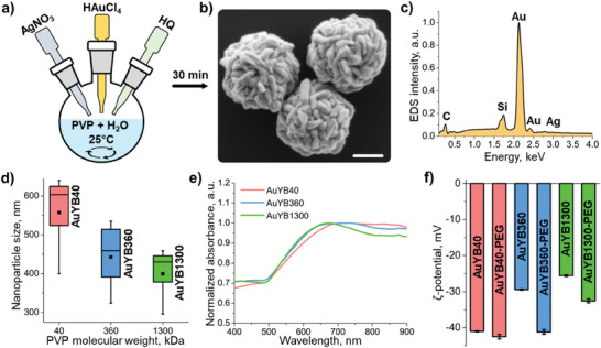
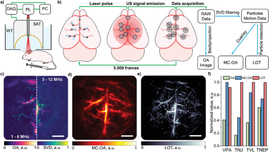
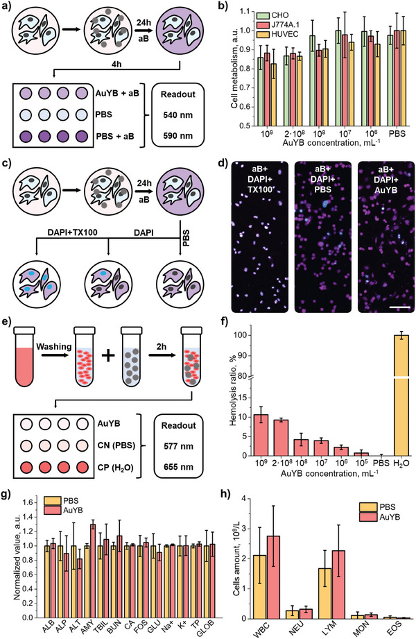
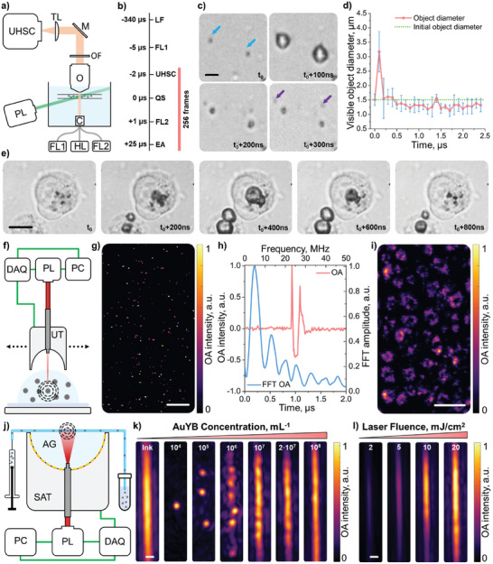
Localization optoacoustic tomography (LOT) has recently emerged as a transformative super-resolution technique breaking through the acoustic diffraction limit in deep-tissue optoacoustic (OA) imaging via individual localization and tracking of particles in the bloodstream. However, strong light absorption in red blood cells has previously restricted per-particle OA detection to relatively large microparticles, ≈5 µm in diameter. Herein, it is demonstrated that submicron-sized porous gold nanoparticles, ≈600 nm in diameter, can be individually detected for noninvasive super-resolution imaging with LOT. Ultra-high-speed bright-field microscopy revealed that these nanoparticles generate microscopic plasmonic vapor bubbles, significantly enhancing opto-acoustic energy conversion through a nano-to-micro size transformation. Comprehensive in vitro and in vivo tests further demonstrated the biocompatibility and biosafety of the particles. By reducing the detectable particle size by an order of magnitude, nanoLOT enables microangiographic imaging with a significantly reduced risk of embolisms from particle aggregation and opens new avenues to visualize how nanoparticles reach vascular and potentially extravascular targets. The performance of nanoLOT for non-invasive imaging of microvascular networks in the murine brain anticipates new insights into neurovascular coupling mechanisms and longitudinal microcirculatory changes associated with neurodegenerative diseases.
局部光声层析成像(LOT)最近已成为一种变革性的超分辨率技术,通过对血流中粒子的个体定位和跟踪,突破了深层组织光声(OA)成像中的声学衍射极限。然而,红细胞中的强光吸收以前将每个粒子的OA检测限制在相对较大的微粒上,直径约为5微米。在此证明,直径约600纳米的亚微米级多孔金纳米粒子可以通过LOT进行个体检测,用于无创超分辨率成像。超高速明场显微镜显示,这些纳米粒子会产生微观等离子体气泡,通过纳米到微米的尺寸转变显著增强光声能量转换。全面的体外和体内测试进一步证明了这些粒子的生物相容性和生物安全性。通过将可检测的粒子尺寸减小一个数量级,纳米LOT实现了微血管造影成像,显著降低了粒子聚集导致栓塞的风险,并为可视化纳米粒子如何到达血管及潜在的血管外靶点开辟了新途径。纳米LOT对小鼠大脑微血管网络进行无创成像的性能有望为神经血管耦合机制以及与神经退行性疾病相关的纵向微循环变化带来新的见解。