Beckman Institute for Advanced Science and Technology, University of Illinois Urbana-Champaign, Urbana, IL, USA.
Department of Electrical and Computer Engineering, University of Illinois Urbana-Champaign, Urbana, IL, USA.
Nat Commun. 2024 Apr 4;15(1):2932. doi: 10.1038/s41467-024-47154-2.
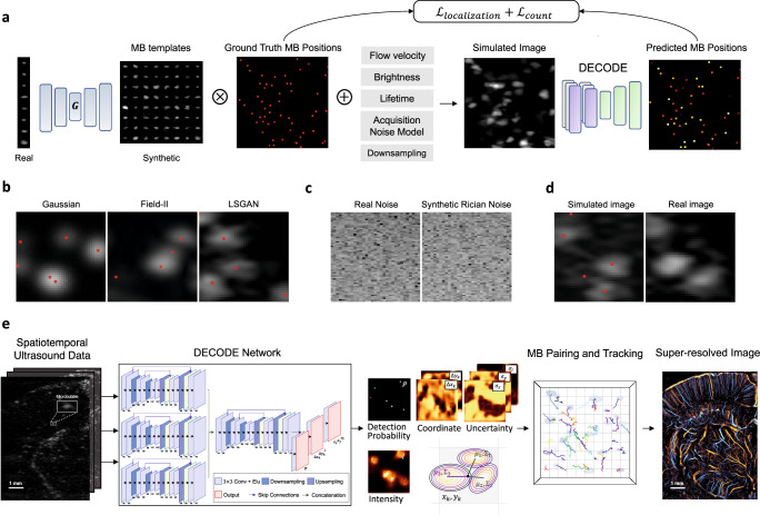
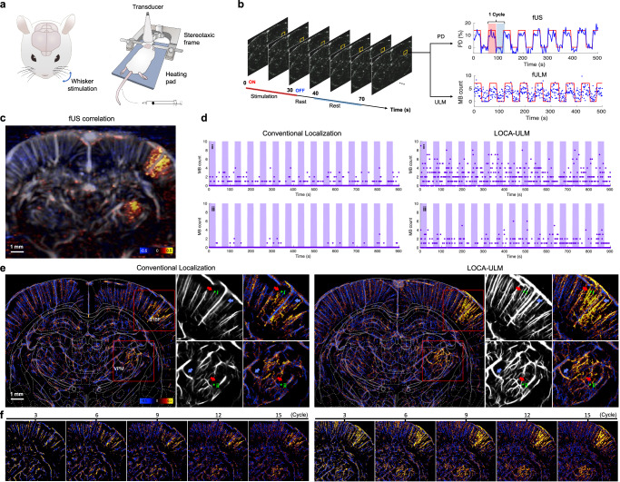
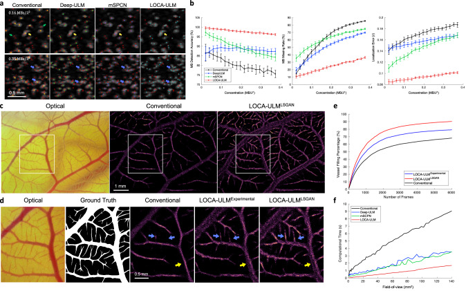
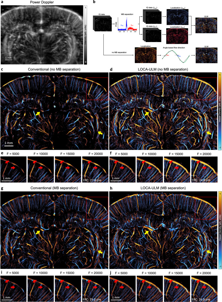
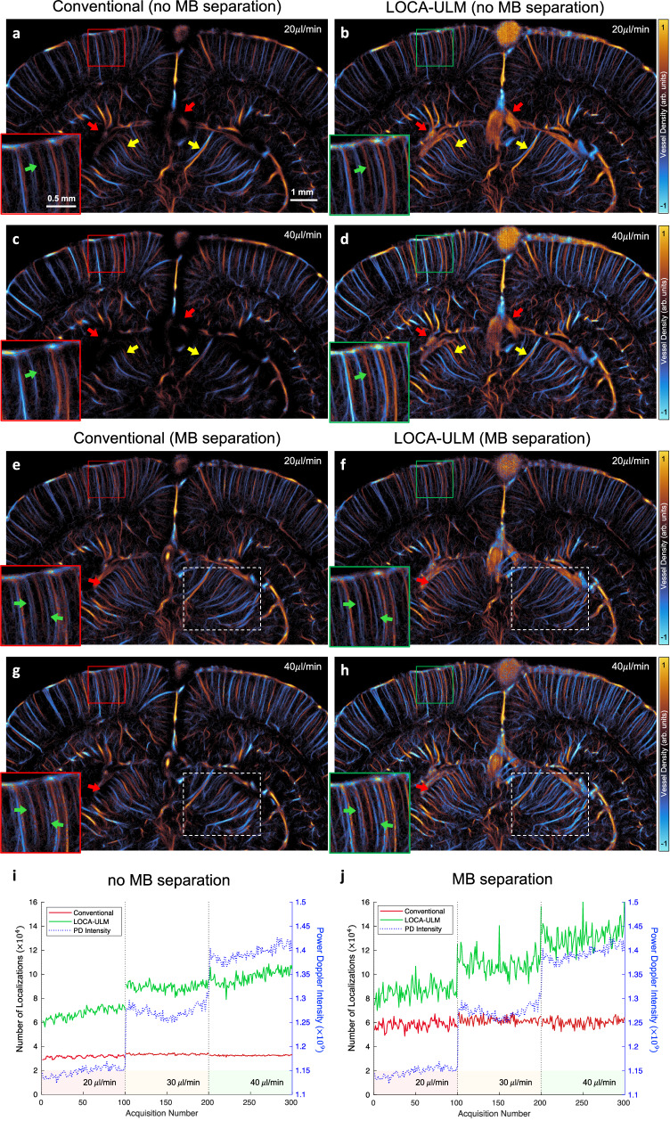
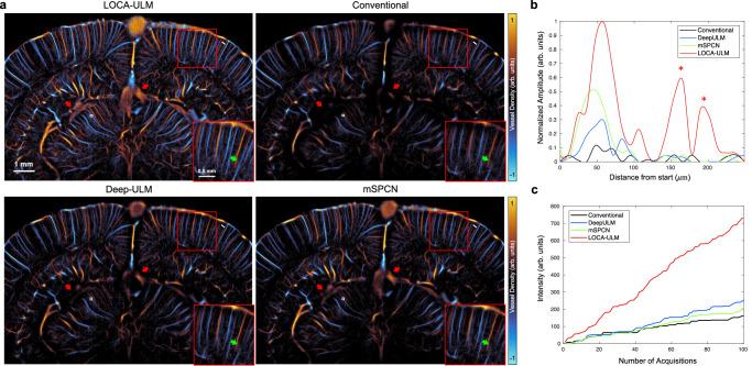
Ultrasound localization microscopy (ULM) enables deep tissue microvascular imaging by localizing and tracking intravenously injected microbubbles circulating in the bloodstream. However, conventional localization techniques require spatially isolated microbubbles, resulting in prolonged imaging time to obtain detailed microvascular maps. Here, we introduce LOcalization with Context Awareness (LOCA)-ULM, a deep learning-based microbubble simulation and localization pipeline designed to enhance localization performance in high microbubble concentrations. In silico, LOCA-ULM enhanced microbubble detection accuracy to 97.8% and reduced the missing rate to 23.8%, outperforming conventional and deep learning-based localization methods up to 17.4% in accuracy and 37.6% in missing rate reduction. In in vivo rat brain imaging, LOCA-ULM revealed dense cerebrovascular networks and spatially adjacent microvessels undetected by conventional ULM. We further demonstrate the superior localization performance of LOCA-ULM in functional ULM (fULM) where LOCA-ULM significantly increased the functional imaging sensitivity of fULM to hemodynamic responses invoked by whisker stimulations in the rat brain.
超声定位显微镜(ULM)通过定位和跟踪在血流中循环的静脉内注射的微泡,实现深层组织微血管成像。然而,传统的定位技术需要空间隔离的微泡,导致获得详细微血管图谱的成像时间延长。在这里,我们介绍了基于深度学习的微泡模拟和定位管道 LOcalization with Context Awareness (LOCA)-ULM,旨在提高高浓度微泡中的定位性能。在计算机模拟中,LOCA-ULM 将微泡检测的准确性提高到 97.8%,并将漏检率降低到 23.8%,在准确性方面优于传统和基于深度学习的定位方法高达 17.4%,在漏检率降低方面高达 37.6%。在大鼠脑的体内成像中,LOCA-ULM 揭示了密集的脑血管网络和空间相邻的微血管,这些在传统的 ULM 中是无法检测到的。我们进一步证明了 LOCA-ULM 在功能 ULM(fULM)中的优越定位性能,在大鼠脑中,LOCA-ULM 显著提高了 fULM 对由胡须刺激引起的血流动力学反应的功能成像灵敏度。