• 文献检索
  • 文档翻译
  • 深度研究
  • 学术资讯
  • Suppr Zotero 插件Zotero 插件
  • 邀请有礼
  • 套餐&价格
  • 历史记录
应用&插件
Suppr Zotero 插件Zotero 插件浏览器插件Mac 客户端Windows 客户端微信小程序
定价
高级版会员购买积分包购买API积分包
服务
文献检索文档翻译深度研究API 文档MCP 服务
关于我们
关于 Suppr公司介绍联系我们用户协议隐私条款
关注我们

Suppr 超能文献

核心技术专利:CN118964589B侵权必究
粤ICP备2023148730 号-1Suppr @ 2026

文献检索

告别复杂PubMed语法,用中文像聊天一样搜索,搜遍4000万医学文献。AI智能推荐,让科研检索更轻松。

立即免费搜索

文件翻译

保留排版,准确专业,支持PDF/Word/PPT等文件格式,支持 12+语言互译。

免费翻译文档

深度研究

AI帮你快速写综述,25分钟生成高质量综述,智能提取关键信息,辅助科研写作。

立即免费体验

亚细胞器分辨率下蛋白质聚集的荧光寿命辅助探测

Fluorescence Lifetime-Assisted Probing of Protein Aggregation with sub-Organellar Resolution.

作者信息

Gupta Karnika, Maddison Daniel C, Melo Eduardo P, da Costa Ana Rosa M, Avezov Edward

机构信息

UK Dementia Research Institute at University of Cambridge, Department of Clinical Neurosciences, Cambridge, UK.

Centre of Marine Sciences (CCMAR/CIMAR LA), Campus de Gambelas, Universidade do Algarve, Faro, Portugal.

出版信息

Bio Protoc. 2024 Oct 5;14(19):e5080. doi: 10.21769/BioProtoc.5080.

DOI:10.21769/BioProtoc.5080
PMID:39399587
原文链接:https://pmc.ncbi.nlm.nih.gov/articles/PMC11470378/
Abstract

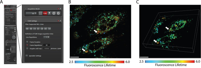
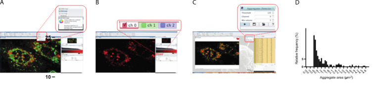
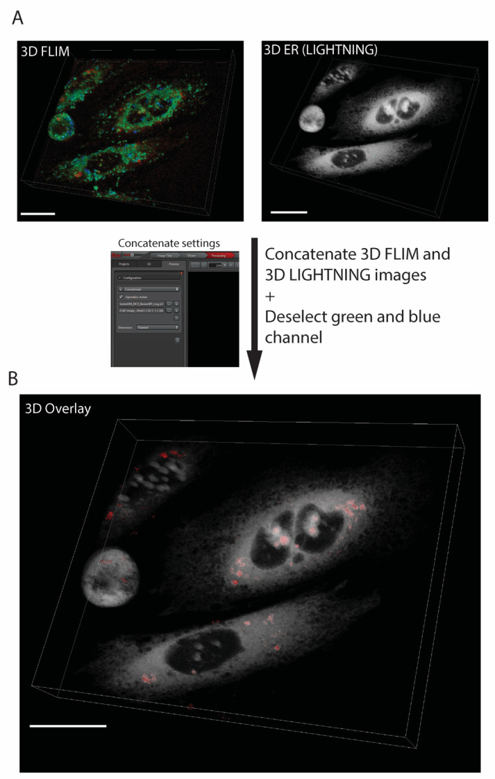
Protein misfolding fuels multiple neurodegenerative diseases, but existing techniques lack the resolution to pinpoint the location and physical properties of aggregates within living cells. Our protocol describes high-resolution confocal and fluorescent lifetime microscopy (Fast 3D FLIM) of an aggregation probing system. This system involves a metastable HaloTag protein (HT-aggr) labeled with P1 solvatochromic fluorophore, which can be targeted to subcellular compartments. This strategy allows to distinguish between aggregated and folded probe species, since P1 fluorophore changes its lifetime depending on the hydrophobicity of its microenvironment. The probe is not fluorescence intensity-dependent, overcoming issues related to intensity-based measurements of labeled proteins, such as control of probe quantity due to differences in expression or photobleaching of a proportion of the fluorophore population. Our approach reports on the performance of the machinery dealing with aggregation-prone substrates and thus opens doors to studying proteostasis and its role in neurodegenerative diseases. Key features • Aggregation state: Tracks aggregate formation and disaggregation with pulse-chase experiments • Sub-organellar resolution: Pinpoints and allows control of aggregate location within the cell, exceeding traditional techniques • Quantitative analysis: Measures aggregate load through image analysis • Methodology: • Metastable HaloTag variant labeling with a solvatochromic small-molecule reporter ligand • High-resolution confocal microscopy coupled with FLIM for aggregate identification and localization • Image analysis for aggregate quantification and distribution within the ER • Pulse-chase experiments to track aggregates.

摘要

蛋白质错误折叠引发多种神经退行性疾病,但现有技术缺乏分辨率来精确确定活细胞内聚集体的位置和物理特性。我们的方案描述了一种聚集探测系统的高分辨率共聚焦和荧光寿命显微镜技术(快速3D荧光寿命成像)。该系统涉及一种用P1溶剂化显色荧光团标记的亚稳态卤代标签蛋白(HT-聚集体),它可以靶向亚细胞区室。这种策略能够区分聚集态和折叠态的探针种类,因为P1荧光团会根据其微环境的疏水性改变其寿命。该探针不依赖荧光强度,克服了与基于强度的标记蛋白测量相关的问题, 例如由于表达差异或部分荧光团群体的光漂白而导致的探针数量控制问题。我们的方法报告了处理易聚集底物的机制的性能,从而为研究蛋白质稳态及其在神经退行性疾病中的作用打开了大门。关键特性 • 聚集状态:通过脉冲追踪实验跟踪聚集体的形成和解聚 • 亚细胞器分辨率:精确确定并控制细胞内聚集体的位置,超越传统技术 • 定量分析:通过图像分析测量聚集体负载 • 方法: • 用溶剂化显色小分子报告配体标记亚稳态卤代标签变体 • 高分辨率共聚焦显微镜与荧光寿命成像相结合用于聚集体鉴定和定位 • 图像分析用于聚集体定量和在内质网中的分布 • 脉冲追踪实验跟踪聚集体

https://cdn.ncbi.nlm.nih.gov/pmc/blobs/9801/11470378/ec759a4efba4/BioProtoc-14-20-5080-v001.jpg
https://cdn.ncbi.nlm.nih.gov/pmc/blobs/9801/11470378/4b163a98f047/BioProtoc-14-20-5080-g001.jpg
https://cdn.ncbi.nlm.nih.gov/pmc/blobs/9801/11470378/ddcc3ef3c434/BioProtoc-14-20-5080-g002.jpg
https://cdn.ncbi.nlm.nih.gov/pmc/blobs/9801/11470378/2e1d3eeba43d/BioProtoc-14-20-5080-g003.jpg
https://cdn.ncbi.nlm.nih.gov/pmc/blobs/9801/11470378/ec759a4efba4/BioProtoc-14-20-5080-v001.jpg

相似文献

1
Fluorescence Lifetime-Assisted Probing of Protein Aggregation with sub-Organellar Resolution.亚细胞器分辨率下蛋白质聚集的荧光寿命辅助探测
Bio Protoc. 2024 Oct 5;14(19):e5080. doi: 10.21769/BioProtoc.5080.
2
Fluorescence lifetime imaging microscopy: fundamentals and advances in instrumentation, analysis, and applications.荧光寿命成像显微镜:仪器、分析和应用的基本原理及进展。
J Biomed Opt. 2020 May;25(7):1-43. doi: 10.1117/1.JBO.25.7.071203.
3
Fluorescence Lifetime Imaging Probes for Cell-Based Measurements of Enzyme Activity.用于基于细胞的酶活性测量的荧光寿命成像探针。
Methods Mol Biol. 2022;2394:133-162. doi: 10.1007/978-1-0716-1811-0_9.
4
A Solvatochromic Fluorescent Probe Reveals Polarity Heterogeneity upon Protein Aggregation in Cells.一种溶致变色荧光探针揭示了细胞中蛋白质聚集时的极性不均匀性。
Angew Chem Int Ed Engl. 2021 Dec 1;60(49):25865-25871. doi: 10.1002/anie.202107943. Epub 2021 Nov 2.
5
Stress-induced protein disaggregation in the endoplasmic reticulum catalysed by BiP.BiP 催化内质网中的应激诱导蛋白解聚。
Nat Commun. 2022 May 6;13(1):2501. doi: 10.1038/s41467-022-30238-2.
6
Quantitative comparison of different fluorescent protein couples for fast FRET-FLIM acquisition.不同荧光蛋白偶联物用于快速 FRET-FLIM 获取的定量比较。
Biophys J. 2009 Oct 21;97(8):2368-76. doi: 10.1016/j.bpj.2009.07.044.
7
Image analysis for denoising full-field frequency-domain fluorescence lifetime images.用于全场频域荧光寿命图像去噪的图像分析
J Microsc. 2009 Aug;235(2):221-37. doi: 10.1111/j.1365-2818.2009.03212.x.
8
Characterization of Amyloid Structures in Aging C. Elegans Using Fluorescence Lifetime Imaging.利用荧光寿命成像技术对衰老秀丽隐杆线虫中的淀粉样蛋白结构进行表征。
J Vis Exp. 2020 Mar 27(157). doi: 10.3791/61004.
9
Visualizing the Multistep Process of Protein Aggregation in Live Cells.可视化活细胞中蛋白质聚集的多步骤过程。
Acc Chem Res. 2022 Feb 1;55(3):381-390. doi: 10.1021/acs.accounts.1c00648. Epub 2022 Jan 18.
10
Fluorescence correlation spectroscopy and fluorescence lifetime imaging microscopy.荧光相关光谱法和荧光寿命成像显微镜技术。
Nephron Exp Nephrol. 2006;103(2):e41-9. doi: 10.1159/000090615. Epub 2006 Mar 10.

本文引用的文献

1
Endoplasmic reticulum morphology regulation by RTN4 modulates neuronal regeneration by curbing luminal transport.内质网形态调节通过抑制腔运输调节神经元再生。
Cell Rep. 2024 Jul 23;43(7):114357. doi: 10.1016/j.celrep.2024.114357. Epub 2024 Jul 1.
2
Luminal transport through intact endoplasmic reticulum limits the magnitude of localized Ca signals.内质网腔运输通过完整的内质网限制局部 Ca 信号的幅度。
Proc Natl Acad Sci U S A. 2024 Mar 26;121(13):e2312172121. doi: 10.1073/pnas.2312172121. Epub 2024 Mar 19.
3
Molecular pathology of neurodegenerative diseases by cryo-EM of amyloids.
淀粉样变 cryo-EM 在神经退行性疾病的分子病理学中的应用。
Nature. 2023 Sep;621(7980):701-710. doi: 10.1038/s41586-023-06437-2. Epub 2023 Sep 27.
4
Stress-induced protein disaggregation in the endoplasmic reticulum catalysed by BiP.BiP 催化内质网中的应激诱导蛋白解聚。
Nat Commun. 2022 May 6;13(1):2501. doi: 10.1038/s41467-022-30238-2.
5
Folding or holding?-Hsp70 and Hsp90 chaperoning of misfolded proteins in neurodegenerative disease.折叠还是持留?——热休克蛋白 70 和 90 在神经退行性疾病中对错误折叠蛋白的伴侣作用。
J Biol Chem. 2022 May;298(5):101905. doi: 10.1016/j.jbc.2022.101905. Epub 2022 Apr 6.
6
The proteostasis network and its decline in ageing.蛋白质稳态网络及其在衰老过程中的衰退。
Nat Rev Mol Cell Biol. 2019 Jul;20(7):421-435. doi: 10.1038/s41580-019-0101-y.
7
Single particle trajectories reveal active endoplasmic reticulum luminal flow.单颗粒轨迹揭示了内质网腔的活跃流动。
Nat Cell Biol. 2018 Oct;20(10):1118-1125. doi: 10.1038/s41556-018-0192-2. Epub 2018 Sep 17.
8
AgHalo: A Facile Fluorogenic Sensor to Detect Drug-Induced Proteome Stress.AgHalo:一种用于检测药物诱导的蛋白质组应激的简便荧光传感器。
Angew Chem Int Ed Engl. 2017 Jul 17;56(30):8672-8676. doi: 10.1002/anie.201702417. Epub 2017 Jun 19.
9
Protein misfolding in the endoplasmic reticulum as a conduit to human disease.内质网中蛋白质的错误折叠作为通向人类疾病的途径。
Nature. 2016 Jan 21;529(7586):326-35. doi: 10.1038/nature17041.
10
Icy: an open bioimage informatics platform for extended reproducible research.Icy:一个开放的生物影像信息学平台,用于扩展可重复研究。
Nat Methods. 2012 Jun 28;9(7):690-6. doi: 10.1038/nmeth.2075.