Jansen Theodorus J P, Tokgöz Sevilay, Buitinga Mijke, van Lith Sanne A M, Joosten Lieke, Frielink Cathelijne, Smeets Esther M M, Stommel Martijn W J, van der Kolk Marion B, de Galan Bastiaan E, Brom Maarten, Boss Marti, Gotthardt Martin
Department of Medical Imaging, Radboud University Medical Center, Nijmegen, The Netherlands.
Nutrition and Movement Sciences, Maastricht University, Maastricht, The Netherlands.
EJNMMI Res. 2024 Oct 15;14(1):96. doi: 10.1186/s13550-024-01159-6.
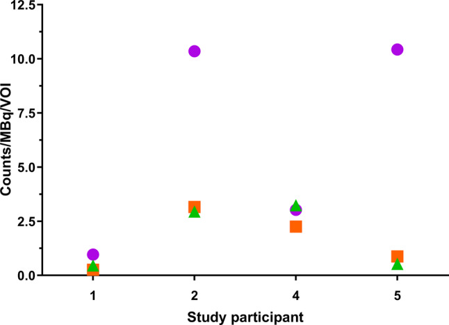
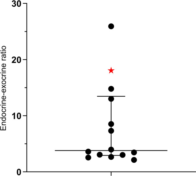
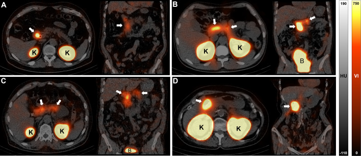
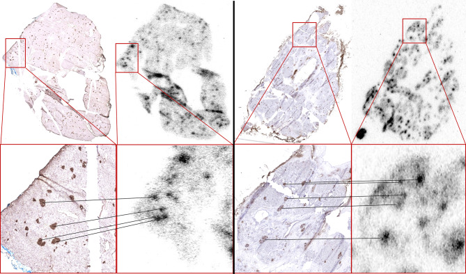
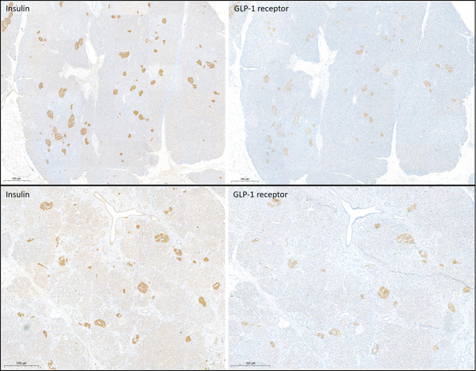
Estimation of beta cell mass is currently restricted to evaluating pancreatic tissue samples, which provides limited information. A non-invasive imaging technique that reliably quantifies beta cell mass enables monitoring of changes of beta cell mass during the progression of diabetes mellitus and may contribute to monitoring of therapy effectiveness. We assessed the specificity of radiolabelled exendin for beta cell mass quantification in humans. Fourteen adults with pancreas tumours were injected with In-labeled exendin-4 prior to pancreatic resection. In resected pancreas tissue, endocrine-exocrine ratios of tracer uptake were determined by digital autoradiography and accumulation of In-labeled exendin-4 was compared to insulin and GLP-1 receptor staining. Of four participants, abdominal single photon emission computed tomography/computed tomography (SPECT/CT) images were acquired to quantify pancreatic uptake in vivo RESULTS: Tracer uptake was predominantly present in the endocrine pancreas (endocrine-exocrine ratio: 3.6 [2.8-10.8]. Tracer accumulation showed overlap with insulin-positive regions, which overlapped with GLP-1 receptor positive areas. SPECT imaging showed pancreatic uptake of radiolabelled exendin in three participants.
Radiolabelled exendin specifically accumulates in the islets of Langerhans in human pancreas tissue. The clear overlap between regions positive for insulin and the GLP-1 receptor substantiate the beta cell specificity of the tracer. Radiolabelled exendin is therefore a valuable imaging agent for human beta cell mass quantification and has the potential to be used for a range of applications, including improvement of diabetes treatment by assessment of the effects of current and novel diabetes therapies on the beta cell mass.
ClinicalTrials.gov NCT03889496, registered 26,032,019, URL https://clinicaltrials.gov/study/NCT03889496?term=NCT03889496 .
gov NCT04733508, registered 02022021, URL https://clinicaltrials.gov/study/NCT04733508 .
目前,β细胞量的评估仅限于对胰腺组织样本进行检测,所提供的信息有限。一种能够可靠地对β细胞量进行定量分析的非侵入性成像技术,可用于监测糖尿病进展过程中β细胞量的变化,并有助于监测治疗效果。我们评估了放射性标记的艾塞那肽在人体β细胞量定量分析中的特异性。14例患有胰腺肿瘤的成年人在胰腺切除术前注射了铟标记的艾塞那肽-4。在切除的胰腺组织中,通过数字放射自显影法测定示踪剂摄取的内分泌-外分泌比率,并将铟标记的艾塞那肽-4的积聚情况与胰岛素和胰高血糖素样肽-1受体染色结果进行比较。对4名参与者进行了腹部单光子发射计算机断层扫描/计算机断层扫描(SPECT/CT)成像,以在体内定量胰腺摄取情况。结果:示踪剂摄取主要存在于内分泌胰腺中(内分泌-外分泌比率:3.6[2.8 - 10.8])。示踪剂积聚与胰岛素阳性区域有重叠,而胰岛素阳性区域又与胰高血糖素样肽-1受体阳性区域重叠。SPECT成像显示3名参与者的胰腺摄取了放射性标记的艾塞那肽。
放射性标记的艾塞那肽特异性地积聚在人体胰腺组织的胰岛中。胰岛素阳性区域与胰高血糖素样肽-1受体阳性区域之间明显的重叠证实了该示踪剂对β细胞的特异性。因此,放射性标记的艾塞那肽是一种用于人体β细胞量定量分析的有价值的成像剂,并且有潜力用于一系列应用,包括通过评估当前和新型糖尿病疗法对β细胞量的影响来改善糖尿病治疗。
ClinicalTrials.gov NCT03889496,于2019年3月26日注册,网址https://clinicaltrials.gov/study/NCT03889496?term=NCT03889496 。
gov NCT04733508,于2021年2月2日注册,网址https://clinicaltrials.gov/study/NCT04733508 。