Department of Radiation Oncology, University of California, Los Angeles, CA, USA.
Division of Nuclear Medicine and Molecular Imaging, Department of Radiology and Radiological Science, Johns Hopkins University School of Medicine, Baltimore, MD, USA.
Sci Rep. 2024 Oct 18;14(1):24411. doi: 10.1038/s41598-024-75589-6.
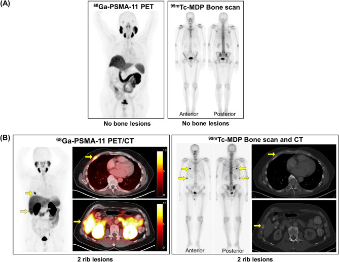
For prostate cancer patients who experience biochemical progression during androgen deprivation therapy (ADT), prostate-specific membrane antigen positron emission tomography/computed tomography (PSMA PET/CT) has not been prospectively compared to planar bone scan plus CT. This was a single-arm, head-to-head, prospective phase II trial (NCT04928820) designed to enroll 102 men with prostate cancer who experienced biochemical progression (rising prostate-specific antigen [PSA] ≥ 1 ng/mL) during ADT. All patients received 68Ga-PSMA-11 PET/CT and 99mTc-MDP planar bone scans. Each scan was interpreted by three central independent readers. The primary endpoint was the per-patient bone metastasis detection rate of PSMA PET/CT versus planar bone scan and CT. Secondary endpoints compared the number of bone metastases detected per patient and the inter-reader agreement of each imaging modality. Twenty-two men were enrolled between July 2021 and June 2022. Due to slow accrual following approval of PSMA PET radiotracers in the U.S. and a lack of a statistical signal between the two imaging modalities on interim analysis, this trial was closed early on October 2022. Median PSA was 8.5 ng/mL (interquartile range: 1.6-77.6). There was 100% agreement between the two scans. Six patients (27%) had negative findings and 16 patients (73%) had positive findings on both scans. PSMA PET/CT and bone scan plus CT detected an equal number of bone lesions for 14 patients (64%), PSMA PET/CT detected more bone lesions for six patients (27%), and bone scan plus CT detected more bone lesions for two patients (9.1%) (p = 0.092). The inter-reader agreement rates of PSMA PET/CT and bone scan plus CT were 96% and 82%, respectively (p = 0.25). In men with biochemical progression during ADT, 68Ga-PSMA-11 PET/CT and 99mTc-MDP planar bone scan plus CT had identical bone metastasis detection rates. Bone scan plus CT can continue to serve as a cost-effective and readily accessible restaging modality in patients with biochemical progression. ClinicalTrials.gov NCT04928820. Registered 16/06/2021.
对于雄激素剥夺治疗(ADT)期间发生生化进展的前列腺癌患者,前列腺特异性膜抗原正电子发射断层扫描/计算机断层扫描(PSMA PET/CT)尚未与平面骨扫描加 CT 进行前瞻性比较。这是一项单臂、头对头、前瞻性 II 期试验(NCT04928820),旨在招募 102 名在 ADT 期间发生生化进展(前列腺特异性抗原 [PSA] 升高≥1ng/mL)的前列腺癌患者。所有患者均接受 68Ga-PSMA-11 PET/CT 和 99mTc-MDP 平面骨扫描。每位患者的扫描均由三位独立的中央读者进行解读。主要终点是 PSMA PET/CT 与平面骨扫描和 CT 的每位患者骨转移检测率。次要终点比较了每位患者检测到的骨转移数量和每种影像学方式的读者间一致性。2021 年 7 月至 2022 年 6 月期间共招募了 22 名男性。由于在美国批准 PSMA PET 放射性示踪剂后入组速度缓慢,以及中期分析中两种影像学方式之间缺乏统计学信号,该试验于 2022 年 10 月提前关闭。中位 PSA 为 8.5ng/mL(四分位距:1.6-77.6)。两种扫描结果完全一致。6 名患者(27%)两种扫描均为阴性,16 名患者(73%)两种扫描均为阳性。PSMA PET/CT 和骨扫描加 CT 分别为 14 名患者(64%)检测到相同数量的骨病变,为 6 名患者(27%)检测到更多的骨病变,为 2 名患者(9.1%)检测到更多的骨病变(p=0.092)。PSMA PET/CT 和骨扫描加 CT 的读者间一致性率分别为 96%和 82%(p=0.25)。在 ADT 期间发生生化进展的男性中,68Ga-PSMA-11 PET/CT 和 99mTc-MDP 平面骨扫描加 CT 的骨转移检测率相同。骨扫描加 CT 可继续作为生化进展患者具有成本效益且易于获得的再分期方式。ClinicalTrials.gov NCT04928820。注册于 2021 年 6 月 16 日。