Cheng Chia-Hsin, Guan Yi, Chiplunkar Vidhi P, Mortazavi Farzad, Medalla Maria L, Sullivan Kimberly, O'Callaghan James P, Koo Bang-Bon, Kelly Kimberly A, Michalovicz Lindsay T
Chobanian & Avedisian School of Medicine, Boston University, Boston, MA, USA.
School of Public Health, Boston University, Boston, MA, USA.
Brain Behav Immun Health. 2024 Sep 30;42:100878. doi: 10.1016/j.bbih.2024.100878. eCollection 2024 Dec.
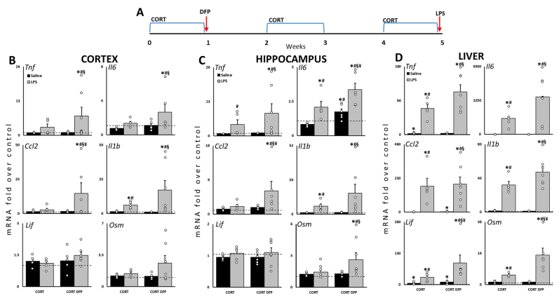
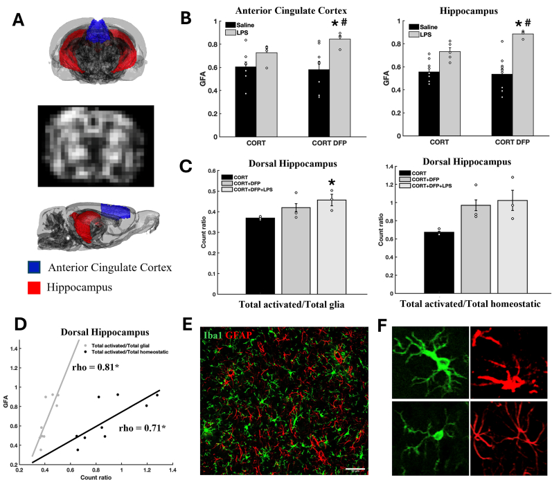
Gulf War Illness (GWI) is a disorder experienced by many veterans of the 1991 Gulf War, with symptoms including fatigue, chronic pain, respiratory and memory problems. Exposure to toxic chemicals during the war, such as oil well fire smoke, pesticides, physiological stress, and nerve agents, is thought to have triggered abnormal neuroinflammatory responses that contribute to GWI. Previous studies have examined the acute effects of combined physiological stress and chemical exposures using GWI rodent models and presented findings related to neuroinflammation and changes in diffusion magnetic resonance imaging (MRI) measures, suggesting a neuroimmune basis for GWI. In the current study, using MRI, cytokine mRNA expression, and immunohistological analyses of brain tissues, we examined the brain structure and immune function of a chronic rat model of GWI. Our data showed that a combination of long-term corticosterone treatment (to mimic high physiological stress) and diisopropyl fluorophosphate exposure (to mimic sarin exposure) primed the response to subsequent systemic immune challenge with lipopolysaccharide resulting in elevations of multiple cytokine mRNAs, an increased activated glial population, and disrupted brain microstructure in the cingulate cortex and hippocampus compared to control groups. Our findings support the critical role of neuroinflammation, dysregulated glial activation, and their relationship to disrupted brain microstructural integrity in the pathophysiology of GWI and highlight the unique consequences of long-term combined exposures on brain biochemistry and structural connectivity.
海湾战争综合征(GWI)是许多1991年海湾战争退伍军人所经历的一种疾病,其症状包括疲劳、慢性疼痛、呼吸和记忆问题。人们认为,战争期间接触有毒化学物质,如油井火灾烟雾、杀虫剂、生理应激和神经毒剂,引发了异常的神经炎症反应,从而导致了海湾战争综合征。先前的研究使用海湾战争综合征啮齿动物模型研究了生理应激和化学暴露联合作用的急性影响,并呈现了与神经炎症和扩散磁共振成像(MRI)测量变化相关的研究结果,提示海湾战争综合征存在神经免疫基础。在当前的研究中,我们使用MRI、细胞因子mRNA表达以及脑组织的免疫组织学分析,对海湾战争综合征慢性大鼠模型的脑结构和免疫功能进行了研究。我们的数据显示,长期给予皮质酮治疗(以模拟高生理应激)和暴露于二异丙基氟磷酸酯(以模拟沙林暴露)相结合,引发了对随后脂多糖全身免疫挑战的反应,导致多种细胞因子mRNA水平升高、活化胶质细胞数量增加,与对照组相比,扣带回皮质和海马体的脑微结构遭到破坏。我们的研究结果支持了神经炎症、胶质细胞激活失调及其与海湾战争综合征病理生理学中脑微结构完整性破坏之间的关系在其中所起的关键作用,并突出了长期联合暴露对脑生物化学和结构连通性的独特影响。