Sanap Arun, Yadav Anita, Choudhary Amruta, Kamath Anusha, Bhimgade Prajakta, Patokar Gauri, Moharana Bishnupriya
Department of Obstetrics and Gynaecology, All India Institute of Medical Sciences, Nagpur, Maharashtra, India.
J Family Reprod Health. 2024 Sep;18(3):197-199. doi: 10.18502/jfrh.v18i3.16662.
To describe the clinical and radio-pathological features of suture granuloma, an inflammatory response to retained suture material that primarily affects non-absorbable sutures.
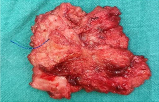
We report a case of a 26-year-old female presenting with painful swelling at a caesarean section scar, previously excised for similar complaints. Physical examination revealed a solid soft tissue mass on the scar. Magnetic resonance imaging (MRI) identified a 2x2 cm lesion in the right abdominal wall, suggestive of suture granuloma. Surgical excision revealed prolene suture material within the granulomatous tissue. Histopathology confirmed foreign body reaction.
Recurrence post-prior excision underscores the importance of complete granuloma removal. Differential diagnoses included scar endometriosis and inflammatory lesions. Suture granulomas, though rare, require consideration in scar-related swelling. Collaboration between specialties ensures accurate diagnosis and management.
描述缝线肉芽肿的临床和放射病理特征,缝线肉芽肿是对留存缝线材料的一种炎症反应,主要影响不可吸收缝线。
我们报告一例26岁女性,其剖宫产瘢痕处出现疼痛性肿胀,该瘢痕此前因类似症状已切除过。体格检查发现瘢痕处有一个实性软组织肿块。磁共振成像(MRI)显示右腹壁有一个2×2厘米的病变,提示缝线肉芽肿。手术切除显示肉芽肿组织内有聚丙烯缝线材料。组织病理学证实为异物反应。
先前切除后复发凸显了彻底清除肉芽肿的重要性。鉴别诊断包括瘢痕子宫内膜异位症和炎性病变。缝线肉芽肿虽罕见,但在瘢痕相关肿胀中需予以考虑。多专科协作可确保准确诊断和治疗。