European Retinoblastoma Imaging Collaboration (ERIC), Amsterdam, The Netherlands.
Cancer Center Amsterdam, Imaging and Biomarkers, Amsterdam, The Netherlands.
Sci Rep. 2024 Oct 23;14(1):25103. doi: 10.1038/s41598-024-76933-6.
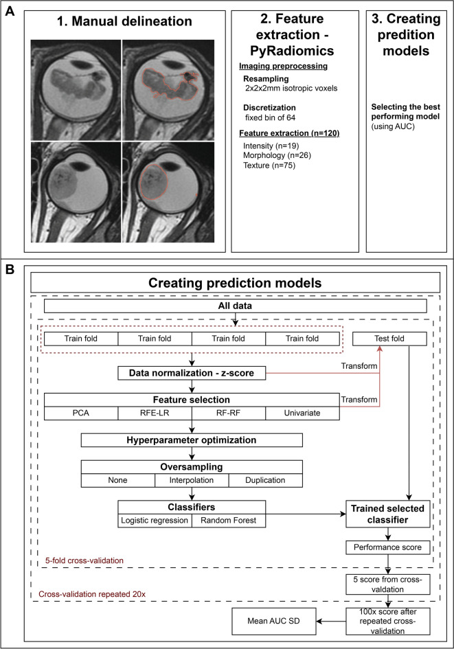
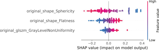
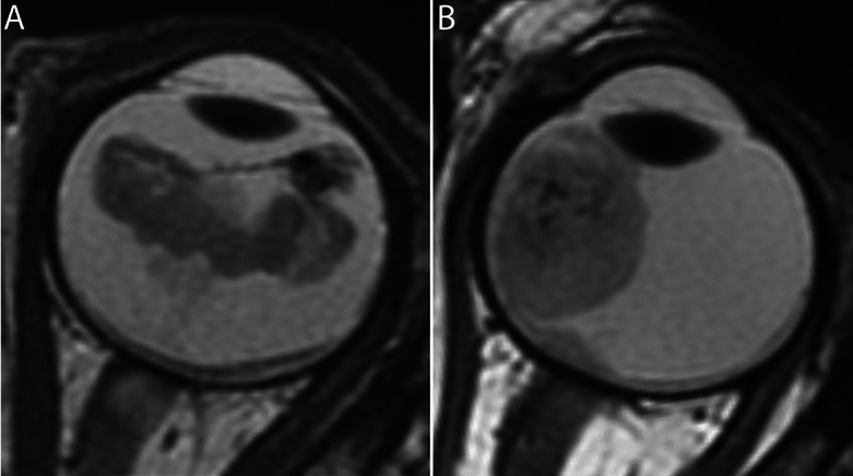
MYCN-amplified RB1 wild-type (MYCNRB1) retinoblastoma is a rare and aggressive subtype, often resistant to standard therapies. Identifying unique MRI features is crucial for diagnosing this subtype, as biopsy is not recommended. This study aimed to differentiate MYCNRB1 from the most prevalent RB1 retinoblastoma using pretreatment MRI and radiomics. Ninety-eight unilateral retinoblastoma patients (19 MYCN cases and 79 matched controls) were included. Tumors on T2-weighted MR images were manually delineated and validated by experienced radiologists. Radiomics analysis extracted 120 features per tumor. Several combinations of feature selection methods, oversampling techniques and machine learning (ML) classifiers were evaluated in a repeated fivefold cross-validation machine learning pipeline to yield the best-performing prediction model for MYCN. The best model used univariate feature selection, data oversampling (duplicating MYCN cases), and logistic regression classifier, achieving a mean AUC of 0.78 (SD 0.12). SHAP analysis highlighted lower sphericity, higher flatness, and greater gray-level heterogeneity as predictive for MYCNRB1 status, yielding an AUC of 0.81 (SD 0.11). This study shows the potential of MRI-based radiomics to distinguish MYCNRB1 and RB1 retinoblastoma subtypes.
MYCN 扩增 RB1 野生型(MYCNRB1)视网膜母细胞瘤是一种罕见且侵袭性的亚型,通常对标准治疗有抗性。确定独特的 MRI 特征对于诊断这种亚型至关重要,因为不建议进行活检。本研究旨在使用预处理 MRI 和放射组学来区分 MYCNRB1 和最常见的 RB1 视网膜母细胞瘤。纳入了 98 例单侧视网膜母细胞瘤患者(19 例 MYCN 病例和 79 例匹配对照)。在 T2 加权 MR 图像上手动勾画肿瘤,并由经验丰富的放射科医生进行验证。放射组学分析从每个肿瘤中提取了 120 个特征。在重复的五折交叉验证机器学习管道中评估了几种特征选择方法、过采样技术和机器学习(ML)分类器的组合,以产生用于预测 MYCN 的性能最佳的预测模型。最佳模型使用单变量特征选择、数据过采样(复制 MYCN 病例)和逻辑回归分类器,平均 AUC 为 0.78(SD 0.12)。SHAP 分析突出了较低的球形度、较高的平坦度和更大的灰度异质性作为预测 MYCNRB1 状态的特征,AUC 为 0.81(SD 0.11)。本研究表明基于 MRI 的放射组学在区分 MYCNRB1 和 RB1 视网膜母细胞瘤亚型方面具有潜力。