Pfefferle Susanne, Schweizer Michaela, Hartmann Kristin, Berger Julia, Nörz Dominik, Emmerich Petra, von Possel Ronald, Giersch Katja, Pflüger Lisa Sophie, Bernreuther Christian, Glatzel Markus, Krasemann Susanne, Brehm Thomas Theo, Schulze Zur Wisch Julian, Fischer Nicole, Schmiedel Stefan, Aepfelbacher Martin, Lütgehetmann Marc
Institute for Medical Microbiology, Virology and Hygiene, University Medical Center Hamburg-Eppendorf, 20246, Hamburg, Germany.
Morphology and Electron Microscopy Core Facility, Center for Molecular Neurobiology (ZMNH), University Medical Center Hamburg-Eppendorf, 20251, Hamburg, Germany.
Heliyon. 2024 Oct 4;10(19):e38873. doi: 10.1016/j.heliyon.2024.e38873. eCollection 2024 Oct 15.
We report prolonged mpox (>14 weeks) in a patient with HIV complicated by deep tissue MPXV infection despite two courses of tecovirimat treatment.
MPXV-DNA levels in lesional swabs, blood and tissue were quantified by qPCR. Anti-MPXV antibodies were analyzed by IF and VNT. Infectivity was assessed by virus isolation. Sequencing was performed to assess for tecovirimat resistance mutations and quantitative results were obtained by digital SNP PCR (A288P).
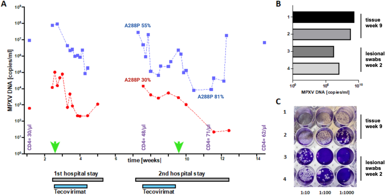
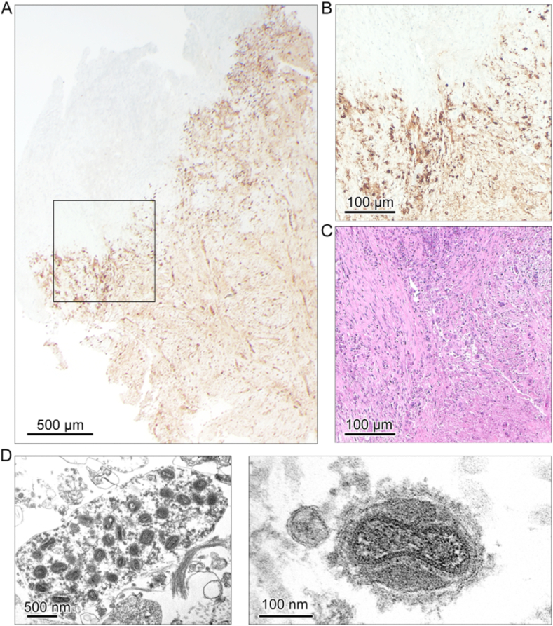
The patient's clinical condition improved significantly during both tecovirimat treatment courses (each 14 days), yet we observed persistent MPXV-DNA in lesions accompanied by viremia (mean 1.4 × 10 copies/ml) for >14 weeks. A deep tissue infection driven by MPXV complicated the clinical course (week 9). Presence of infectious virus within the tissue and high infectious titers (>10 PFU/ml) were observed. The VP37 protein sequence revealed A288P substitutions. Digital PCR showed 1 % and less abundance (A288P) during first treatment course (blood and swabs), with increasing proportion during second course (week 8-9; 28 % in blood and swabs), however the mutation was absent in samples from deep tissue infection and MPXV isolates (week 9) indicating compartimentalization. Morphological fully enveloped MPXV partices visualized by TEM in necrotic areas suggesting tecovirimat treatment failure in the deep tissue compartment.
Our data provide evidence that Tecovirimat treatment selects for compartimentalized viral mutations (A288P). While the patient clinically benefited from repeated tecovirimat course, emergence of viral muations and deep tissue infection emphasizes the challenge and importance of infectious disease monitoring in mpox patient management.
我们报告了一名感染艾滋病毒的患者出现了持续时间较长的猴痘(超过14周),尽管接受了两个疗程的特考韦瑞治疗,但仍并发了深部组织猴痘病毒感染。
通过qPCR对病变拭子、血液和组织中的猴痘病毒DNA水平进行定量。通过免疫荧光和病毒中和试验分析抗猴痘病毒抗体。通过病毒分离评估传染性。进行测序以评估特考韦瑞耐药突变,并通过数字单核苷酸多态性PCR(A288P)获得定量结果。
在两个特考韦瑞治疗疗程(各14天)期间,患者的临床状况有显著改善,但我们观察到病变中猴痘病毒DNA持续存在,并伴有病毒血症(平均1.4×10拷贝/毫升)超过14周。由猴痘病毒驱动的深部组织感染使临床病程复杂化(第9周)。在组织中观察到有传染性病毒存在且传染性滴度较高(>10 PFU/毫升)。VP37蛋白序列显示存在A288P替代。数字PCR显示在第一个疗程(血液和拭子)期间A288P的丰度为1%及以下,在第二个疗程(第8 - 9周;血液和拭子中为28%)期间比例增加,然而在深部组织感染样本和猴痘病毒分离株(第9周)中未发现该突变,表明存在分区现象。通过透射电子显微镜在坏死区域观察到形态完整的包膜猴痘病毒颗粒,提示特考韦瑞治疗在深部组织区域失败。
我们的数据提供了证据,表明特考韦瑞治疗会选择出分区化的病毒突变(A288P)。虽然患者从重复的特考韦瑞疗程中临床获益,但病毒突变和深部组织感染的出现强调了在猴痘患者管理中传染病监测的挑战和重要性。