• 文献检索
  • 文档翻译
  • 深度研究
  • 学术资讯
  • Suppr Zotero 插件Zotero 插件
  • 邀请有礼
  • 套餐&价格
  • 历史记录
应用&插件
Suppr Zotero 插件Zotero 插件浏览器插件Mac 客户端Windows 客户端微信小程序
定价
高级版会员购买积分包购买API积分包
服务
文献检索文档翻译深度研究API 文档MCP 服务
关于我们
关于 Suppr公司介绍联系我们用户协议隐私条款
关注我们

Suppr 超能文献

核心技术专利:CN118964589B侵权必究
粤ICP备2023148730 号-1Suppr @ 2026

文献检索

告别复杂PubMed语法,用中文像聊天一样搜索,搜遍4000万医学文献。AI智能推荐,让科研检索更轻松。

立即免费搜索

文件翻译

保留排版,准确专业,支持PDF/Word/PPT等文件格式,支持 12+语言互译。

免费翻译文档

深度研究

AI帮你快速写综述,25分钟生成高质量综述,智能提取关键信息,辅助科研写作。

立即免费体验

使用三维岛状绒毛膜容积(3D-ICRV)技术对适于胎龄胎儿和早发性胎儿生长受限胎儿的岛状发育进行超声评估。

Ultrasound assessment of insular development in adequate-for-gestational-age fetuses and fetuses with early-onset fetal growth restriction using 3D-ICRV technology.

作者信息

Xue Jinfeng, Xue Jinluan, Ru Yanhui, Zhang Ge, Yin Hong, Liu Dequan

机构信息

School of Medical Imaging, Shandong Second Medical University, Weifang, China.

Department of Ultrasound, Shandong Provincial Maternal and Child Health Care Hospital, Jinan, China.

出版信息

Front Med (Lausanne). 2024 Oct 9;11:1393115. doi: 10.3389/fmed.2024.1393115. eCollection 2024.

DOI:10.3389/fmed.2024.1393115
PMID:39444811
原文链接:https://pmc.ncbi.nlm.nih.gov/articles/PMC11496279/
Abstract

OBJECTIVE

This study aimed to evaluate the growth trajectory of the insula in adequate-for-gestational-age (AGA) and early-onset fetal growth restriction (FGR) fetuses and analyze the difference between the two groups using three-dimensional inversion crytal and realistic vue technique (3D-ICRV).

METHODS

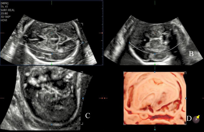
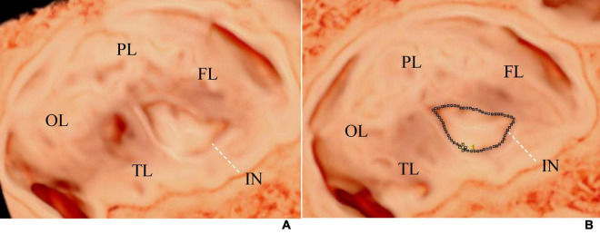
Singleton pregnant women, with a gestational age ranging from 20 to 32 weeks, who underwent routine examinations at Shandong Maternal and Child Care Hospital between March 2023 and December 2023 were included. The participants were divided into two groups: the FGR and AGA fetuses. Three-dimensional volumes were obtained using transabdominal ultrasound in the transverse section of the fetal hypothalamus based on different gestational ages. 3D-ICRV rendering technology was used for 3D imaging of the fetal insula. Volumes with a clear display of the insula were selected. We observed the morphology of the insula, and measured the area and circumference of the insula. By evaluating the growth trajectory of the insula in AGA and FGR fetuses, differences in insular development between the two groups were compared.

RESULTS

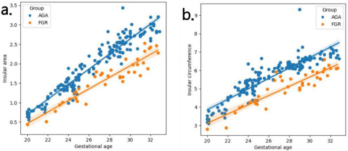
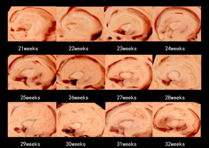
Overall, 203 participants were included in this study, with 164 and 39 in the AGA and FGR groups, respectively. The 3D volumes were successfully acquired, and the area and circumference of the insula were measured using 3D-ICRV imaging technology. We found that as gestational age increased, the area and circumference of the insula gradually increased and showed positive correlations with the gestational age, with no significant changes in morphology. The growth rate of insular area and insular circumference in the FGR group is slower than that in the AGA group (insular area: 0.15 vs 0.19 cm / week, insular circumference: 0.25 vs 0.28 cm / week). The area and circumference of the insula in the FGR group were significantly different from those in the AGA group (insular area: = 0.003, insular circumference: = 0.004).

CONCLUSION

The measured values of the insula using 3D-ICRV identify the differences in insular development between the FGR and AGA fetuses. The findings of this study have important implications for the prenatal evaluation of cortical development and maturity in FGR fetuses and further clinical consultation and management.

摘要

目的

本研究旨在评估适于胎龄(AGA)和早发型胎儿生长受限(FGR)胎儿脑岛的生长轨迹,并使用三维反转晶体和逼真视图技术(3D-ICRV)分析两组之间的差异。

方法

纳入2023年3月至2023年12月期间在山东省妇幼保健院接受常规检查、孕周为20至32周的单胎孕妇。参与者分为两组:FGR胎儿组和AGA胎儿组。基于不同孕周,经腹超声在下丘脑横切面获取三维容积数据。采用3D-ICRV渲染技术对胎儿脑岛进行三维成像。选择脑岛显示清晰的容积数据。观察脑岛形态,测量脑岛面积和周长。通过评估AGA和FGR胎儿脑岛的生长轨迹,比较两组脑岛发育的差异。

结果

本研究共纳入203名参与者,AGA组164例,FGR组39例。成功获取三维容积数据,并使用3D-ICRV成像技术测量脑岛面积和周长。我们发现,随着孕周增加,脑岛面积和周长逐渐增大,且与孕周呈正相关,形态无明显变化。FGR组脑岛面积和脑岛周长的生长速度慢于AGA组(脑岛面积:0.15 vs 0.19 cm²/周,脑岛周长:0.25 vs 0.28 cm/周)。FGR组脑岛面积和周长与AGA组有显著差异(脑岛面积:P = 0.003,脑岛周长:P = 0.004)。

结论

使用3D-ICRV测量的脑岛值可识别FGR和AGA胎儿脑岛发育的差异。本研究结果对FGR胎儿皮质发育和成熟度的产前评估以及进一步的临床咨询和管理具有重要意义。

https://cdn.ncbi.nlm.nih.gov/pmc/blobs/9496/11496279/e7b6fe8d893c/fmed-11-1393115-g004.jpg
https://cdn.ncbi.nlm.nih.gov/pmc/blobs/9496/11496279/8252e2f93b15/fmed-11-1393115-g001.jpg
https://cdn.ncbi.nlm.nih.gov/pmc/blobs/9496/11496279/946de25110d9/fmed-11-1393115-g002.jpg
https://cdn.ncbi.nlm.nih.gov/pmc/blobs/9496/11496279/1944aae90225/fmed-11-1393115-g003.jpg
https://cdn.ncbi.nlm.nih.gov/pmc/blobs/9496/11496279/e7b6fe8d893c/fmed-11-1393115-g004.jpg

相似文献

1
Ultrasound assessment of insular development in adequate-for-gestational-age fetuses and fetuses with early-onset fetal growth restriction using 3D-ICRV technology.使用三维岛状绒毛膜容积(3D-ICRV)技术对适于胎龄胎儿和早发性胎儿生长受限胎儿的岛状发育进行超声评估。
Front Med (Lausanne). 2024 Oct 9;11:1393115. doi: 10.3389/fmed.2024.1393115. eCollection 2024.
2
Midline structures and cortical development in late-onset fetal growth restriction according to Doppler status: prospective study.根据多普勒状态评估晚发型胎儿生长受限的中线结构和皮质发育:前瞻性研究。
Ultrasound Obstet Gynecol. 2024 Aug;64(2):228-235. doi: 10.1002/uog.27598. Epub 2024 Jul 7.
3
Fetal insular measurements in pregnancy with estimated fetal weight <10th centile and childhood neurodevelopmental outcomes.胎儿岛叶在估计胎儿体重<第 10 百分位数与儿童神经发育结局的孕期测量。
Am J Obstet Gynecol. 2024 Jan;230(1):85.e1-85.e15. doi: 10.1016/j.ajog.2023.07.009. Epub 2023 Jul 11.
4
Sonographic demonstration of the sulci and gyri on the convex surface in normal fetuses using 3D-ICRV rendering technology.应用 3D-ICRV 渲染技术在正常胎儿凸面上显示脑沟和脑回。
Ultraschall Med. 2023 Dec;44(6):e284-e295. doi: 10.1055/a-2122-6182. Epub 2023 Jul 4.
5
Fetal neurosonography detects differences in cortical development and corpus callosum in late-onset small fetuses.胎儿神经超声检查可检测出晚发型小胎儿皮质发育和胼胝体的差异。
Ultrasound Obstet Gynecol. 2021 Jul;58(1):42-47. doi: 10.1002/uog.23592.
6
Measurement of the Brain Volume/Liver Volume Ratio by Three-Dimensional MRI in Appropriate-for-Gestational Age Fetuses and Those With Fetal Growth Restriction.通过三维磁共振成像测量适于胎龄胎儿和胎儿生长受限胎儿的脑体积/肝体积比
J Magn Reson Imaging. 2021 Dec;54(6):1796-1801. doi: 10.1002/jmri.27792. Epub 2021 Jun 21.
7
Neurological development may be accelerated in growth-restricted fetuses: a 4D ultrasound study.生长受限胎儿的神经发育可能加快:一项 4D 超声研究。
J Perinat Med. 2019 May 27;47(4):429-433. doi: 10.1515/jpm-2018-0379.
8
Reduced adipose tissue in growth-restricted fetuses using quantitative analysis of magnetic resonance images.生长受限胎儿磁共振图像定量分析脂肪组织减少。
Eur Radiol. 2023 Dec;33(12):9194-9202. doi: 10.1007/s00330-023-09855-y. Epub 2023 Jun 30.
9
Evaluation of fetal cardiac function in fetal growth restriction via fetal HQ analysis based on two-dimensional STI.基于二维 STI 的胎儿 HQ 分析对胎儿生长受限胎儿心功能的评价。
J Obstet Gynaecol Res. 2023 Jun;49(6):1514-1524. doi: 10.1111/jog.15631. Epub 2023 Mar 9.
10
Three-Dimensional Power Doppler Evaluation of Cerebral Vascular Blood Flow: A Novel Tool in the Assessment of Fetal Growth Restriction.三维能量多普勒评估脑血管血流:胎儿生长受限评估中的一种新工具。
J Ultrasound Med. 2018 Jan;37(1):139-147. doi: 10.1002/jum.14323. Epub 2017 Jul 14.

引用本文的文献

1
Detecting microcephaly and macrocephaly from ultrasound images using artificial intelligence.使用人工智能从超声图像中检测小头畸形和巨头畸形。
BMC Med Imaging. 2025 May 26;25(1):183. doi: 10.1186/s12880-025-01709-x.
2
Risk of congenital anomalies associated with psychotropic medications: a review of neonatal reports in the FDA adverse event reporting System (FAERS).与精神药物相关的先天性异常风险:对美国食品药品监督管理局不良事件报告系统(FAERS)中新生儿报告的综述。
Arch Womens Ment Health. 2024 Dec 23. doi: 10.1007/s00737-024-01540-3.

本文引用的文献

1
Fetal insular measurements in pregnancy with estimated fetal weight <10th centile and childhood neurodevelopmental outcomes.胎儿岛叶在估计胎儿体重<第 10 百分位数与儿童神经发育结局的孕期测量。
Am J Obstet Gynecol. 2024 Jan;230(1):85.e1-85.e15. doi: 10.1016/j.ajog.2023.07.009. Epub 2023 Jul 11.
2
Sonographic demonstration of the sulci and gyri on the convex surface in normal fetuses using 3D-ICRV rendering technology.应用 3D-ICRV 渲染技术在正常胎儿凸面上显示脑沟和脑回。
Ultraschall Med. 2023 Dec;44(6):e284-e295. doi: 10.1055/a-2122-6182. Epub 2023 Jul 4.
3
Three-Dimensional Crystal Vue Imaging technology assessment of Sylvian fissures at 20-32+6 weeks' normal gestation.
20至32⁺⁶周正常妊娠时大脑外侧裂的三维晶体视野成像技术评估
Eur Radiol. 2023 Apr;33(4):2358-2366. doi: 10.1007/s00330-022-09253-w. Epub 2022 Nov 17.
4
Prenatal diagnosis of midgut volvulus using two-dimensional and three-dimensional ultrasound.应用二维和三维超声对中肠扭转进行产前诊断。
Am J Transl Res. 2022 Mar 15;14(3):1859-1867. eCollection 2022.
5
Brain charts for the human lifespan.人类寿命的大脑图谱。
Nature. 2022 Apr;604(7906):525-533. doi: 10.1038/s41586-022-04554-y. Epub 2022 Apr 6.
6
The role of hypoxia in stem cell regulation of the central nervous system: From embryonic development to adult proliferation.缺氧在中枢神经系统干细胞调控中的作用:从胚胎发育到成年增殖。
CNS Neurosci Ther. 2021 Dec;27(12):1446-1457. doi: 10.1111/cns.13754.
7
Severe early-onset fetal growth restriction: What do we tell the prospective parents?重度早发型胎儿生长受限:我们该如何告知准父母?
Prenat Diagn. 2021 Oct;41(11):1363-1371. doi: 10.1002/pd.6030. Epub 2021 Aug 25.
8
Two-dimensional Speckle tracking echocardiography in Fetal Growth Restriction: a systematic review.二维斑点追踪超声心动图在胎儿生长受限中的应用:一项系统评价
Eur J Obstet Gynecol Reprod Biol. 2020 Nov;254:87-94. doi: 10.1016/j.ejogrb.2020.08.052. Epub 2020 Sep 2.
9
ISUOG Practice Guidelines (updated): sonographic examination of the fetal central nervous system. Part 1: performance of screening examination and indications for targeted neurosonography.国际妇产科超声学会(ISUOG)实践指南(更新版):胎儿中枢神经系统的超声检查。第1部分:筛查检查的实施及针对性神经超声检查的指征
Ultrasound Obstet Gynecol. 2020 Sep;56(3):476-484. doi: 10.1002/uog.22145.
10
Association of Antenatal Corticosteroids and Magnesium Sulfate Therapy With Neurodevelopmental Outcome in Extremely Preterm Children.产前皮质类固醇和硫酸镁治疗与极早产儿神经发育结局的关系。
Obstet Gynecol. 2020 Jun;135(6):1377-1386. doi: 10.1097/AOG.0000000000003882.