• 文献检索
  • 文档翻译
  • 深度研究
  • 学术资讯
  • Suppr Zotero 插件Zotero 插件
  • 邀请有礼
  • 套餐&价格
  • 历史记录
应用&插件
Suppr Zotero 插件Zotero 插件浏览器插件Mac 客户端Windows 客户端微信小程序
定价
高级版会员购买积分包购买API积分包
服务
文献检索文档翻译深度研究API 文档MCP 服务
关于我们
关于 Suppr公司介绍联系我们用户协议隐私条款
关注我们

Suppr 超能文献

核心技术专利:CN118964589B侵权必究
粤ICP备2023148730 号-1Suppr @ 2026

文献检索

告别复杂PubMed语法,用中文像聊天一样搜索,搜遍4000万医学文献。AI智能推荐,让科研检索更轻松。

立即免费搜索

文件翻译

保留排版,准确专业,支持PDF/Word/PPT等文件格式,支持 12+语言互译。

免费翻译文档

深度研究

AI帮你快速写综述,25分钟生成高质量综述,智能提取关键信息,辅助科研写作。

立即免费体验

Pentacam随机森林指数在中国人群中检测亚临床圆锥角膜的作用

The Role of Pentacam Random Forest Index in Detecting Subclinical Keratoconus in a Chinese Cohort.

作者信息

Liu Yan, Zhang Yu, Wang Yuexin, Dong Ruilan, Chen Yueguo

机构信息

Department of Ophthalmology, Peking University Third Hospital, 49 North Garden Road, Haidian District, Beijing 100191, China.

Beijing Key Laboratory of Restoration of Damaged Ocular Nerve, Peking University Third Hospital, Beijing 100191, China.

出版信息

Diagnostics (Basel). 2024 Oct 17;14(20):2304. doi: 10.3390/diagnostics14202304.

DOI:10.3390/diagnostics14202304
PMID:39451627
原文链接:https://pmc.ncbi.nlm.nih.gov/articles/PMC11506929/
Abstract

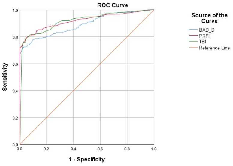
This study aimed to evaluate the diagnostic accuracy of a novel shape index, the Pentacam Random Forest Index (PRFI), in detecting keratoconus (KC), specifically subclinical keratoconus, in Chinese refractive surgery candidates. This prospective cohort study included 856 participants who were divided into four groups based on their tomographic outcomes: the KC group ( = 137), the very asymmetric ectasia (VAE) group ( = 73), the normal cornea group ( = 363) and the tomographically suspected KC (TSK) group ( = 283). The diagnostic performance of PRFI and other widely used indices, including the shape index BAD-D and the combined index TBI, was assessed using receiver operating characteristic (ROC) curve analysis and compared using DeLong's test. The area under the curve (AUC), best cutoff values, and Youden index for each parameter are reported. Additionally, the false-positive rates of BAD-D and PRFI were calculated and compared in "normal corneas". All shape and biomechanical parameters collected in this study were found to be significantly different among the four groups (KC, VAE, TSK, and normal groups; = 0.000). The AUC of PRFI was the highest in detecting any form of KC (including clinical KC eyes and VAE-NT eyes) in Chinese refractive surgery candidates, outperforming the widely used shape index BAD-D (0.919 vs. 0.890, < 0.001). There was no significant difference in performance between the PRFI and the combined TBI index (0.919 vs. 0.916, > 0.05). For detecting subclinical KC eyes (i.e., VAE-NT), the AUC of PRFI was 0.774, which was statistically comparable to TBI (0.774 vs. 0.776, > 0.05), while outperforming BAD-D (0.774 vs. 0.684, < 0.001). The best cutoff values of PRFI for detecting any KC and VAE-NT eyes were determined to be 0.37 and 0.27, respectively. Additionally, PRFI demonstrated a lower false-positive rate than BAD-D (13.8% vs. 43.8%, < 0.001). Notably, the relatively high false-positive rate of BAD-D observed in this study might be attributed to the smaller horizontal corneal diameter in tomographically suspected eyes. The PRFI proved to be a superior shape index compared to BAD-D in detecting any form of keratoconus, including subclinical cases, in Chinese refractive surgery candidates. This finding may be attributed to the relatively small corneas commonly observed in Asians.

摘要

本研究旨在评估一种新型形状指数——Pentacam随机森林指数(PRFI)在中国屈光手术候选者中检测圆锥角膜(KC),特别是亚临床圆锥角膜的诊断准确性。这项前瞻性队列研究纳入了856名参与者,根据他们的断层扫描结果将其分为四组:KC组(n = 137)、极不对称扩张(VAE)组(n = 73)、正常角膜组(n = 363)和断层扫描疑似KC(TSK)组(n = 283)。使用受试者工作特征(ROC)曲线分析评估PRFI和其他广泛使用的指数(包括形状指数BAD - D和综合指数TBI)的诊断性能,并使用德龙检验进行比较。报告了每个参数的曲线下面积(AUC)、最佳截断值和尤登指数。此外,计算并比较了BAD - D和PRFI在“正常角膜”中的假阳性率。本研究收集的所有形状和生物力学参数在四组(KC组、VAE组、TSK组和正常组)之间均存在显著差异(P = 0.000)。在中国屈光手术候选者中,PRFI在检测任何形式的KC(包括临床KC眼和VAE - NT眼)方面的AUC最高,优于广泛使用的形状指数BAD - D(0.919对0.890,P < 0.001)。PRFI与综合TBI指数在性能上无显著差异(0.919对0.916,P > 0.05)。对于检测亚临床KC眼(即VAE - NT),PRFI的AUC为0.774,与TBI在统计学上相当(0.774对0.776,P > 0.05),同时优于BAD - D(0.774对0.684,P < 0.001)。检测任何KC和VAE - NT眼的PRFI最佳截断值分别确定为0.37和0.27。此外,PRFI的假阳性率低于BAD - D(13.8%对43.8%,P < 0.001)。值得注意的是,本研究中观察到的BAD - D相对较高的假阳性率可能归因于断层扫描疑似眼中较小的角膜水平直径。在检测中国屈光手术候选者中的任何形式的圆锥角膜(包括亚临床病例)方面,PRFI被证明是一种优于BAD - D的形状指数。这一发现可能归因于亚洲人常见的相对较小的角膜。

https://cdn.ncbi.nlm.nih.gov/pmc/blobs/e74e/11506929/8b782f68fe15/diagnostics-14-02304-g004.jpg
https://cdn.ncbi.nlm.nih.gov/pmc/blobs/e74e/11506929/18a5f49202df/diagnostics-14-02304-g001.jpg
https://cdn.ncbi.nlm.nih.gov/pmc/blobs/e74e/11506929/c38e513c02d1/diagnostics-14-02304-g002.jpg
https://cdn.ncbi.nlm.nih.gov/pmc/blobs/e74e/11506929/1a0e6326cb3e/diagnostics-14-02304-g003.jpg
https://cdn.ncbi.nlm.nih.gov/pmc/blobs/e74e/11506929/8b782f68fe15/diagnostics-14-02304-g004.jpg

相似文献

1
The Role of Pentacam Random Forest Index in Detecting Subclinical Keratoconus in a Chinese Cohort.Pentacam随机森林指数在中国人群中检测亚临床圆锥角膜的作用
Diagnostics (Basel). 2024 Oct 17;14(20):2304. doi: 10.3390/diagnostics14202304.
2
The Role of Corneal Biomechanics for the Evaluation of Ectasia Patients.角膜生物力学在膨隆患者评估中的作用。
Int J Environ Res Public Health. 2020 Mar 23;17(6):2113. doi: 10.3390/ijerph17062113.
3
Application of a scheimpflug-based biomechanical analyser and tomography in the early detection of subclinical keratoconus in chinese patients.基于 Scheimpflug 技术的生物力学分析和断层成像在中国人亚临床圆锥角膜早期检测中的应用。
BMC Ophthalmol. 2021 Sep 20;21(1):339. doi: 10.1186/s12886-021-02102-2.
4
Comparison of Ectasia Detection in Early Keratoconus Using Scheimpflug-Based Corneal Tomography and Biomechanical Assessments.基于 Scheimpflug 角膜断层成像和生物力学评估的早期圆锥角膜扩张检测比较。
Cornea. 2023 Dec 1;42(12):1528-1535. doi: 10.1097/ICO.0000000000003273. Epub 2023 Mar 27.
5
Enhanced Tomographic Assessment to Detect Corneal Ectasia Based on Artificial Intelligence.基于人工智能的增强断层扫描评估以检测角膜扩张。
Am J Ophthalmol. 2018 Nov;195:223-232. doi: 10.1016/j.ajo.2018.08.005. Epub 2018 Aug 9.
6
New artificial intelligence index based on Scheimpflug corneal tomography to distinguish subclinical keratoconus from healthy corneas.基于 Scheimpflug 角膜断层成像术的新型人工智能指数可区分亚临床圆锥角膜与健康角膜。
J Cataract Refract Surg. 2022 Oct 1;48(10):1168-1174. doi: 10.1097/j.jcrs.0000000000000946.
7
Integration of Scheimpflug-Based Corneal Tomography and Biomechanical Assessments for Enhancing Ectasia Detection.基于Scheimpflug原理的角膜地形图与生物力学评估相结合以增强圆锥角膜检测
J Refract Surg. 2017 Jul 1;33(7):434-443. doi: 10.3928/1081597X-20170426-02.
8
Evaluation of corneal topographic, tomographic and biomechanical indices for detecting clinical and subclinical keratoconus: a comprehensive three-device study.用于检测临床和亚临床圆锥角膜的角膜地形图、断层扫描和生物力学指标评估:一项全面的三设备研究
Int J Ophthalmol. 2021 Feb 18;14(2):228-239. doi: 10.18240/ijo.2021.02.08. eCollection 2021.
9
Comparison of the morphological and biomechanical characteristics of keratoconus, forme fruste keratoconus, and normal corneas.比较圆锥角膜、亚临床圆锥角膜和正常角膜的形态和生物力学特征。
Semin Ophthalmol. 2021 Nov 17;36(8):671-678. doi: 10.1080/08820538.2021.1896752. Epub 2021 Mar 18.
10
Detection of Subclinical Corneal Ectasia Using Corneal Tomographic and Biomechanical Assessments in a Japanese Population.使用角膜断层成像和生物力学评估在日本人群中检测亚临床性角膜扩张。
J Refract Surg. 2019 Jun 1;35(6):383-390. doi: 10.3928/1081597X-20190417-01.

本文引用的文献

1
Combining Spectral-Domain OCT and Air-Puff Tonometry Analysis to Diagnose Keratoconus.联合频域光学相干断层扫描与气吹式眼压测量分析诊断圆锥角膜
J Refract Surg. 2022 Jun;38(6):374-380. doi: 10.3928/1081597X-20220414-02. Epub 2022 Jun 1.
2
New artificial intelligence index based on Scheimpflug corneal tomography to distinguish subclinical keratoconus from healthy corneas.基于 Scheimpflug 角膜断层成像术的新型人工智能指数可区分亚临床圆锥角膜与健康角膜。
J Cataract Refract Surg. 2022 Oct 1;48(10):1168-1174. doi: 10.1097/j.jcrs.0000000000000946.
3
The false positive rates for detecting keratoconus and potential ectatic corneal conditions when evaluating astigmatic eyes with Scheimpflug Technology.
当使用 Scheimpflug 技术评估散光眼时,检测圆锥角膜和潜在扩张性角膜病变的假阳性率。
Eur J Ophthalmol. 2022 Sep;32(5):2532-2546. doi: 10.1177/11206721221081467. Epub 2022 Mar 21.
4
Biomechanics in Keratoconus Diagnosis.圆锥角膜诊断中的生物力学
Curr Eye Res. 2023 Feb;48(2):130-136. doi: 10.1080/02713683.2022.2041042. Epub 2022 Mar 21.
5
Keratoconus: An updated review.圆锥角膜:更新综述。
Cont Lens Anterior Eye. 2022 Jun;45(3):101559. doi: 10.1016/j.clae.2021.101559. Epub 2022 Jan 4.
6
Application of a scheimpflug-based biomechanical analyser and tomography in the early detection of subclinical keratoconus in chinese patients.基于 Scheimpflug 技术的生物力学分析和断层成像在中国人亚临床圆锥角膜早期检测中的应用。
BMC Ophthalmol. 2021 Sep 20;21(1):339. doi: 10.1186/s12886-021-02102-2.
7
Keratoconus: Diagnosis and Staging.圆锥角膜:诊断与分期。
Cornea. 2022 Jan 1;41(1):1-11. doi: 10.1097/ICO.0000000000002781.
8
Comparison of the morphological and biomechanical characteristics of keratoconus, forme fruste keratoconus, and normal corneas.比较圆锥角膜、亚临床圆锥角膜和正常角膜的形态和生物力学特征。
Semin Ophthalmol. 2021 Nov 17;36(8):671-678. doi: 10.1080/08820538.2021.1896752. Epub 2021 Mar 18.
9
Evaluation of corneal topographic, tomographic and biomechanical indices for detecting clinical and subclinical keratoconus: a comprehensive three-device study.用于检测临床和亚临床圆锥角膜的角膜地形图、断层扫描和生物力学指标评估:一项全面的三设备研究
Int J Ophthalmol. 2021 Feb 18;14(2):228-239. doi: 10.18240/ijo.2021.02.08. eCollection 2021.
10
Analysis of OPD-Scan and Pentacam Parameters for Early Keratoconus Detection.分析门诊扫描和 Pentacam 参数,以早期发现圆锥角膜。
Am J Ophthalmol. 2021 Jun;226:235-242. doi: 10.1016/j.ajo.2021.01.012. Epub 2021 Jan 30.