• 文献检索
  • 文档翻译
  • 深度研究
  • 学术资讯
  • Suppr Zotero 插件Zotero 插件
  • 邀请有礼
  • 套餐&价格
  • 历史记录
应用&插件
Suppr Zotero 插件Zotero 插件浏览器插件Mac 客户端Windows 客户端微信小程序
定价
高级版会员购买积分包购买API积分包
服务
文献检索文档翻译深度研究API 文档MCP 服务
关于我们
关于 Suppr公司介绍联系我们用户协议隐私条款
关注我们

Suppr 超能文献

核心技术专利:CN118964589B侵权必究
粤ICP备2023148730 号-1Suppr @ 2026

文献检索

告别复杂PubMed语法,用中文像聊天一样搜索,搜遍4000万医学文献。AI智能推荐,让科研检索更轻松。

立即免费搜索

文件翻译

保留排版,准确专业,支持PDF/Word/PPT等文件格式,支持 12+语言互译。

免费翻译文档

深度研究

AI帮你快速写综述,25分钟生成高质量综述,智能提取关键信息,辅助科研写作。

立即免费体验

增生性糖尿病视网膜病变微环境通过环氧化酶-2/前列腺素 E2 信号通路驱动小胶质细胞极化,促进血管生成和纤维化。

Proliferative Diabetic Retinopathy Microenvironment Drives Microglial Polarization and Promotes Angiogenesis and Fibrosis via Cyclooxygenase-2/Prostaglandin E2 Signaling.

机构信息

Department of Ophthalmology, Juntendo University Urayasu Hospital, 2-1-1 Urayasu, Chiba 279-0021, Japan.

Department of Regenerative Medicine, Graduate School of Medicine, Chiba University, Chiba 263-8522, Japan.

出版信息

Int J Mol Sci. 2024 Oct 21;25(20):11307. doi: 10.3390/ijms252011307.

DOI:10.3390/ijms252011307
PMID:39457089
原文链接:https://pmc.ncbi.nlm.nih.gov/articles/PMC11508523/
Abstract

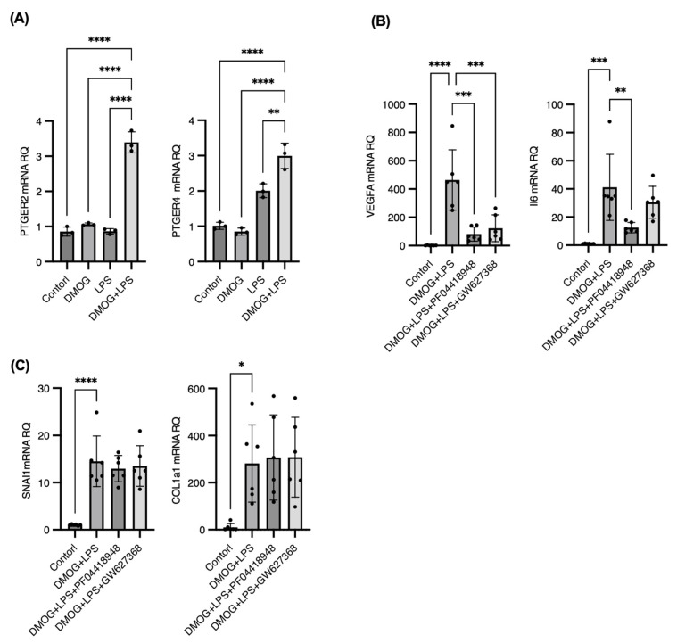
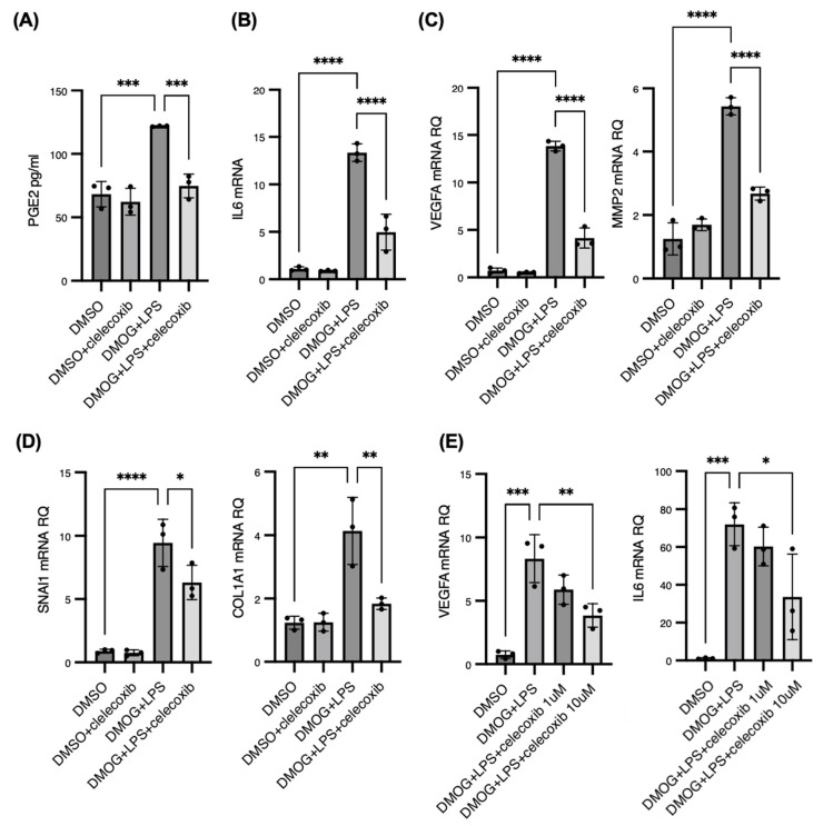
Diabetic retinopathy (DR) is the leading cause of visual impairment, particularly in the proliferative form (proliferative DR [PDR]). The impact of the PDR microenvironment on microglia, which are the resident immune cells in the central nervous system, and the specific pathological changes it may induce remain unclear. This study aimed to investigate the role of microglia in the progression of PDR under hypoxic and inflammatory conditions. We performed a comprehensive gene expression analysis using human-induced pluripotent stem cell-derived microglia under different stimuli (dimethyloxalylglycine (DMOG), lipopolysaccharide (LPS), and DMOG + LPS) to mimic the hypoxic inflammatory environment characteristic of PDR. Principal component analysis revealed distinct gene expression profiles, with 76 genes synergistically upregulated under combined stimulation. Notably, prostaglandin-endoperoxide synthase 2 (encoding cyclooxygenase (COX)-2) exhibited the most pronounced increase, leading to elevated prostaglandin E2 (PGE2) levels and driving pathological angiogenesis and inflammation via the COX-2/PGE2/PGE receptor 2 signaling axis. Additionally, the upregulation of the fibrogenic genes snail family transcriptional repressor 1 and collagen type I alpha 1 chain suggested a role for microglia in fibrosis. These findings underscore the critical involvement of microglia in PDR and suggest that targeting both the angiogenic and fibrotic pathways may present new therapeutic strategies for managing this condition.

摘要

糖尿病性视网膜病变(DR)是视力损害的主要原因,特别是在增生性形式(增生性 DR [PDR])中。PDR 微环境对小胶质细胞的影响,小胶质细胞是中枢神经系统的固有免疫细胞,以及它可能引起的具体病理变化尚不清楚。本研究旨在探讨小胶质细胞在缺氧和炎症条件下 PDR 进展中的作用。我们使用不同刺激(二甲草酰基甘氨酸(DMOG)、脂多糖(LPS)和 DMOG+LPS)下的人诱导多能干细胞衍生小胶质细胞进行了全面的基因表达分析,以模拟 PDR 特征性的缺氧炎症环境。主成分分析显示出明显不同的基因表达谱,76 个基因在联合刺激下协同上调。值得注意的是,前列腺素内过氧化物合酶 2(编码环氧化酶(COX)-2)的表达增加最为明显,导致前列腺素 E2(PGE2)水平升高,并通过 COX-2/PGE2/PGE 受体 2 信号轴驱动病理性血管生成和炎症。此外,成纤维基因 snail 家族转录阻遏物 1 和胶原类型 I alpha 1 链的上调表明小胶质细胞在纤维化中起作用。这些发现强调了小胶质细胞在 PDR 中的关键作用,并表明靶向血管生成和纤维化途径可能为治疗这种疾病提供新的治疗策略。

https://cdn.ncbi.nlm.nih.gov/pmc/blobs/27d2/11508523/e21ffb403444/ijms-25-11307-g006.jpg
https://cdn.ncbi.nlm.nih.gov/pmc/blobs/27d2/11508523/9a7810fc4a6c/ijms-25-11307-g001.jpg
https://cdn.ncbi.nlm.nih.gov/pmc/blobs/27d2/11508523/e94aecb764b7/ijms-25-11307-g002.jpg
https://cdn.ncbi.nlm.nih.gov/pmc/blobs/27d2/11508523/d4d507373e63/ijms-25-11307-g003.jpg
https://cdn.ncbi.nlm.nih.gov/pmc/blobs/27d2/11508523/76d5dba59776/ijms-25-11307-g004.jpg
https://cdn.ncbi.nlm.nih.gov/pmc/blobs/27d2/11508523/5fa5e1eda445/ijms-25-11307-g005.jpg
https://cdn.ncbi.nlm.nih.gov/pmc/blobs/27d2/11508523/e21ffb403444/ijms-25-11307-g006.jpg

相似文献

1
Proliferative Diabetic Retinopathy Microenvironment Drives Microglial Polarization and Promotes Angiogenesis and Fibrosis via Cyclooxygenase-2/Prostaglandin E2 Signaling.增生性糖尿病视网膜病变微环境通过环氧化酶-2/前列腺素 E2 信号通路驱动小胶质细胞极化,促进血管生成和纤维化。
Int J Mol Sci. 2024 Oct 21;25(20):11307. doi: 10.3390/ijms252011307.
2
Differential expression of E-type prostanoid receptors 2 and 4 in microglia stimulated with lipopolysaccharide.脂多糖刺激的小胶质细胞中E型前列腺素受体2和4的差异表达。
J Neuroinflammation. 2017 Jan 5;14(1):3. doi: 10.1186/s12974-016-0780-7.
3
Expression of cyclooxygenase-1/-2, microsomal prostaglandin-E synthase-1 and E-prostanoid receptor 2 and regulation of inflammatory mediators by PGE(2) in the amoeboid microglia in hypoxic postnatal rats and murine BV-2 cells.低氧新生大鼠阿米巴样小胶质细胞和鼠 BV-2 细胞中环氧化酶-1/-2、微粒体前列腺素 E 合酶-1 和 E-前列腺素受体 2 的表达以及 PGE2 对炎症介质的调节。
Neuroscience. 2009 Dec 15;164(3):948-62. doi: 10.1016/j.neuroscience.2009.08.044. Epub 2009 Aug 25.
4
PGE2 Inhibits IL-10 Production via EP2-Mediated β-Arrestin Signaling in Neuroinflammatory Condition.在神经炎症状态下,前列腺素E2通过EP2介导的β-抑制蛋白信号传导抑制白细胞介素-10的产生。
Mol Neurobiol. 2015 Aug;52(1):587-600. doi: 10.1007/s12035-014-8889-0. Epub 2014 Sep 14.
5
Microglia in retinal angiogenesis and diabetic retinopathy.视网膜血管生成和糖尿病视网膜病变中的小胶质细胞。
Angiogenesis. 2024 Aug;27(3):311-331. doi: 10.1007/s10456-024-09911-1. Epub 2024 Apr 2.
6
Prostaglandin E increases the expression of cyclooxygenase-2 in cultured rat microglia.前列腺素 E 增加培养的大鼠小胶质细胞中环氧化酶-2 的表达。
J Neuroimmunol. 2021 Dec 15;361:577724. doi: 10.1016/j.jneuroim.2021.577724. Epub 2021 Sep 23.
7
EP2 receptor signaling pathways regulate classical activation of microglia.EP2 受体信号通路调节小胶质细胞的经典激活。
J Biol Chem. 2013 Mar 29;288(13):9293-302. doi: 10.1074/jbc.M113.455816. Epub 2013 Feb 12.
8
Cyclooxygenase-2 in human and experimental ischemic proliferative retinopathy.环氧化酶-2在人类及实验性缺血性增殖性视网膜病变中的作用
Circulation. 2003 Jul 15;108(2):198-204. doi: 10.1161/01.CIR.0000080735.93327.00. Epub 2003 Jun 23.
9
The microenvironment of proliferative diabetic retinopathy supports lymphatic neovascularization.增殖性糖尿病视网膜病变的微环境支持淋巴血管新生。
J Pathol. 2018 Jun;245(2):172-185. doi: 10.1002/path.5070. Epub 2018 Apr 6.
10
A Systematic Investigation on Complement Pathway Activation in Diabetic Retinopathy.糖尿病性视网膜病变中补体途径激活的系统研究。
Front Immunol. 2020 Feb 11;11:154. doi: 10.3389/fimmu.2020.00154. eCollection 2020.

引用本文的文献

1
CEP78 regulates the proliferation and angiogenesis of endothelial cells by mediating microtubule stabilization via p150glued.CEP78通过p150glued介导微管稳定来调节内皮细胞的增殖和血管生成。
J Mol Histol. 2025 Aug 21;56(5):270. doi: 10.1007/s10735-025-10556-7.
2
EMMPRIN aggravates angiogenesis and blood-retina barrier injury by regulating matrix metalloproteinases in diabetic retinopathy.在糖尿病视网膜病变中,细胞外基质金属蛋白酶诱导因子通过调节基质金属蛋白酶加重血管生成和血视网膜屏障损伤。
Diab Vasc Dis Res. 2025 Mar-Apr;22(2):14791641251324556. doi: 10.1177/14791641251324556. Epub 2025 Apr 18.

本文引用的文献

1
Toll-Like Receptor Signalling Pathways and the Pathogenesis of Retinal Diseases.Toll样受体信号通路与视网膜疾病的发病机制
Front Ophthalmol (Lausanne). 2022 Mar 31;2:850394. doi: 10.3389/fopht.2022.850394. eCollection 2022.
2
Heterotypic macrophages/microglia differentially contribute to retinal ischaemia and neovascularisation.异质性巨噬细胞/小胶质细胞对视网膜缺血和新生血管形成有不同的贡献。
Diabetologia. 2024 Oct;67(10):2329-2345. doi: 10.1007/s00125-024-06215-3. Epub 2024 Jul 8.
3
Microglia in retinal angiogenesis and diabetic retinopathy.
视网膜血管生成和糖尿病视网膜病变中的小胶质细胞。
Angiogenesis. 2024 Aug;27(3):311-331. doi: 10.1007/s10456-024-09911-1. Epub 2024 Apr 2.
4
Microglia-endothelial cross-talk regulates diabetes-induced retinal vascular dysfunction through remodeling inflammatory microenvironment.小胶质细胞与内皮细胞的相互作用通过重塑炎症微环境调节糖尿病诱导的视网膜血管功能障碍。
iScience. 2024 Feb 6;27(3):109145. doi: 10.1016/j.isci.2024.109145. eCollection 2024 Mar 15.
5
Celecoxib/Cyclodextrin Eye Drop Microsuspensions: Evaluation of In Vitro Cytotoxicity and Anti-VEGF Efficacy for Retinal Diseases.塞来昔布/环糊精滴眼液微悬浮液:视网膜疾病的体外细胞毒性和抗血管内皮生长因子疗效评估
Pharmaceutics. 2023 Nov 28;15(12):2689. doi: 10.3390/pharmaceutics15122689.
6
A unique cell population expressing the Epithelial-Mesenchymal Transition-transcription factor Snail moderates microglial and astrocyte injury responses.一种表达上皮-间质转化转录因子Snail的独特细胞群可调节小胶质细胞和星形胶质细胞的损伤反应。
PNAS Nexus. 2023 Oct 12;2(10):pgad334. doi: 10.1093/pnasnexus/pgad334. eCollection 2023 Oct.
7
Persistent diabetic macular edema: Definition, incidence, biomarkers, and treatment methods.持续性糖尿病性黄斑水肿:定义、发生率、生物标志物和治疗方法。
Surv Ophthalmol. 2023 Mar-Apr;68(2):147-174. doi: 10.1016/j.survophthal.2022.11.008. Epub 2022 Nov 24.
8
Deletion of Tgfβ signal in activated microglia prolongs hypoxia-induced retinal neovascularization enhancing Igf1 expression and retinal leukostasis.激活的小胶质细胞中 TGFβ 信号的缺失延长了低氧诱导的视网膜新生血管形成,增强了 Igf1 的表达和视网膜白细胞淤滞。
Glia. 2022 Sep;70(9):1762-1776. doi: 10.1002/glia.24218. Epub 2022 May 25.
9
The role of lipopolysaccharides in diabetic retinopathy.脂多糖在糖尿病性视网膜病变中的作用。
BMC Ophthalmol. 2022 Feb 22;22(1):86. doi: 10.1186/s12886-022-02296-z.
10
Single-Cell Transcriptomics Reveals Novel Role of Microglia in Fibrovascular Membrane of Proliferative Diabetic Retinopathy.单细胞转录组学揭示小胶质细胞在增殖性糖尿病视网膜病变纤维血管膜中的新作用。
Diabetes. 2022 Apr 1;71(4):762-773. doi: 10.2337/db21-0551.