Laboratory of Developmental Biology and Genomics, College of Veterinary Medicine, Interdisciplinary Program for Bioinformatics and Program for Cancer Biology, Seoul National University, Seoul, 08826, Republic of Korea.
Department of New Biology, DGIST, Daegu, 42988, Republic of Korea.
Exp Mol Med. 2024 Oct;56(10):2309-2322. doi: 10.1038/s12276-024-01252-9. Epub 2024 Oct 28.
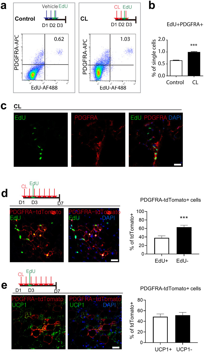
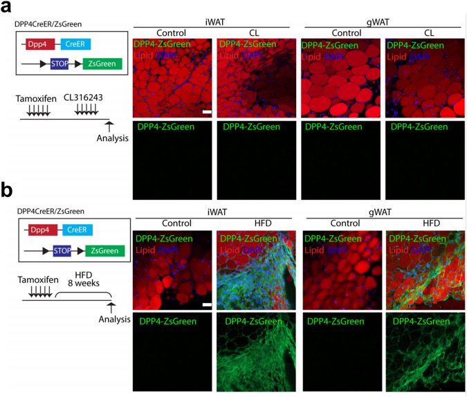
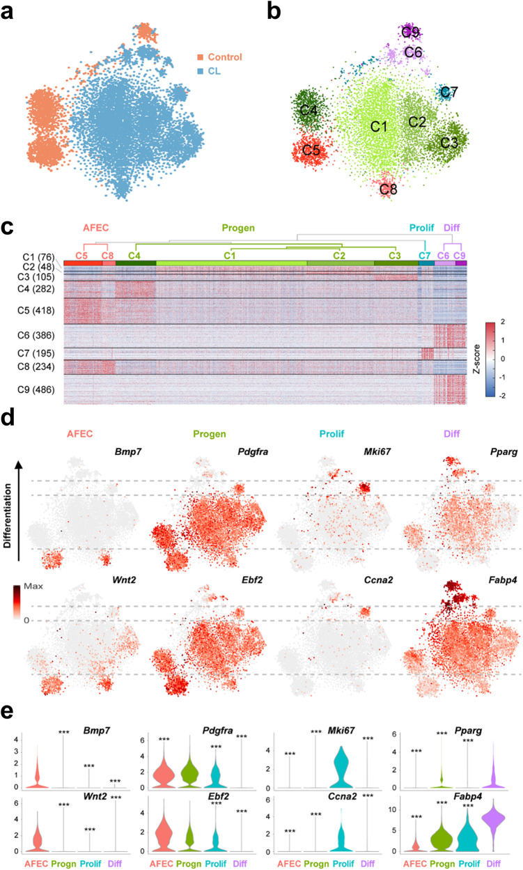
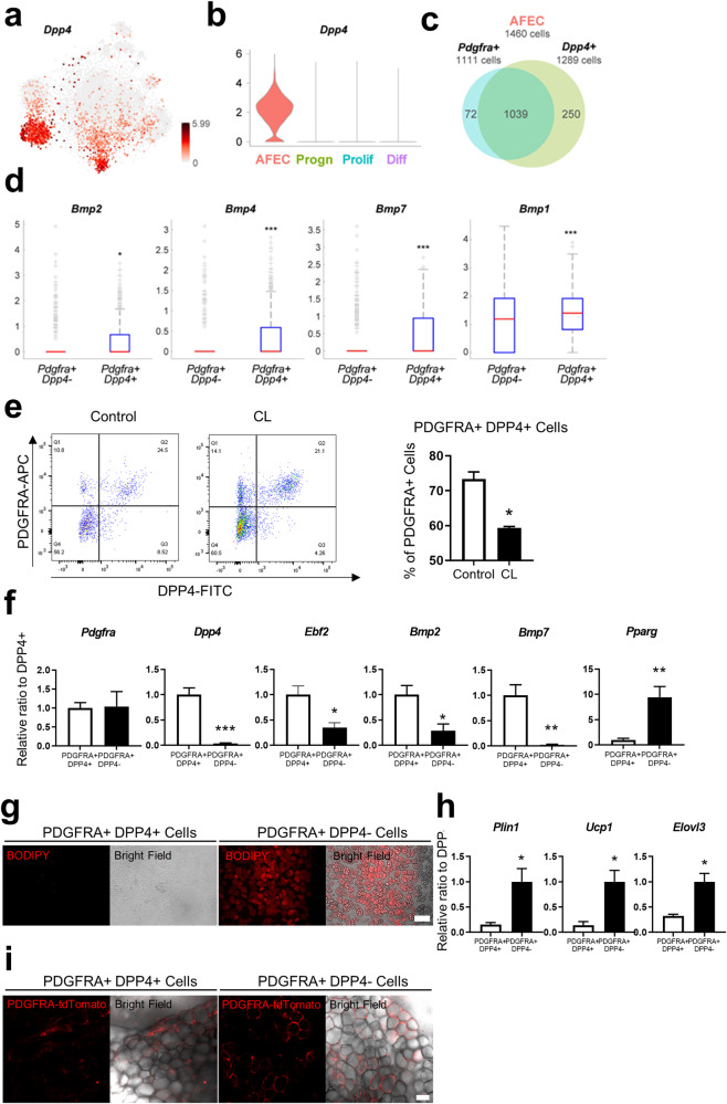
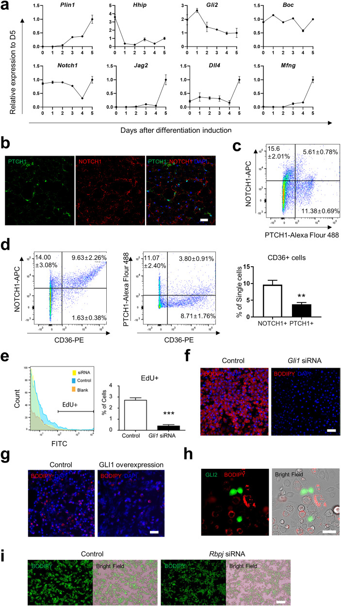
PDGFRA+ cells have been identified as adipocyte stem cells (ASCs) that differentiate into beige adipocytes in white adipose tissue (WAT) following thermogenic stimuli. To elucidate the molecular heterogeneity of ASCs, we conducted single-cell transcriptomic profiling of PDGFRA+ cells isolated from the inguinal WAT (iWAT) of mice treated with the beta3 adrenergic receptor agonist CL316243. Single-cell RNA-seq revealed nine major clusters, which were categorized into four groups: resting, proliferating, differentiating, and adipogenic factor-expressing cells (AFECs). Trajectory analysis revealed sequential activation of molecular pathways, including the Hedgehog and Notch signaling pathways, during beige adipogenesis. AFECs expressed Dpp4 and did not differentiate into adipocytes in culture or after transplantation. Furthermore, genetic lineage tracing studies indicated that DPP4+ cells did not differentiate into adipocytes in iWAT during CL316243-induced beige adipogenesis. However, high-fat diet feeding led to the recruitment of adipocytes from DPP4+ cells in iWAT. Overall, this study improved our understanding of the dynamic molecular basis of beige adipogenesis and the potential contribution of DPP4+ adipocyte lineages to the pathological expansion of WAT during diet-induced obesity.
PDGFRA+ 细胞已被鉴定为脂肪干细胞 (ASCs),它们在白色脂肪组织 (WAT) 中受到产热刺激后可分化为米色脂肪细胞。为了阐明 ASCs 的分子异质性,我们对用β3 肾上腺素能受体激动剂 CL316243 处理的小鼠腹股沟 WAT (iWAT) 中分离的 PDGFRA+ 细胞进行了单细胞转录组谱分析。单细胞 RNA-seq 揭示了九个主要簇,它们分为四组:静止、增殖、分化和脂肪生成因子表达细胞 (AFECs)。轨迹分析显示,在米色脂肪生成过程中,包括 Hedgehog 和 Notch 信号通路在内的分子途径依次被激活。AFECs 表达 Dpp4,在培养或移植后不会分化为脂肪细胞。此外,遗传谱系追踪研究表明,在 CL316243 诱导的米色脂肪生成过程中,DPP4+ 细胞不会分化为 iWAT 中的脂肪细胞。然而,高脂肪饮食喂养导致 iWAT 中的 DPP4+ 细胞募集到脂肪细胞。总的来说,这项研究提高了我们对米色脂肪生成的动态分子基础的理解,以及 DPP4+ 脂肪细胞谱系在饮食诱导肥胖期间 WAT 病理性扩张中的潜在贡献。