Albiña-Palmarola Pablo, Díaz-Peregrino Roberto, Muñoz Sebastian, Lopez Eduardo, Henkes Hans, Mura Jorge
Neuroradiologische Klinik, Klinikum Stuttgart, Stuttgart, Germany.
Department of Neurosurgery, University Hospital Heidelberg, Ruprecht-Karls-University Heidelberg, Heidelberg, Germany.
J Neurosurg Case Lessons. 2024 Oct 28;8(18). doi: 10.3171/CASE24484.
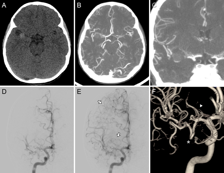
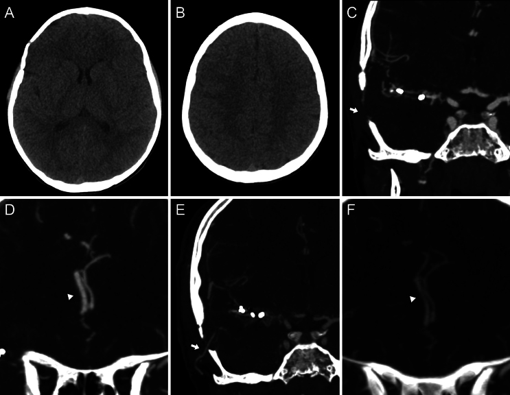
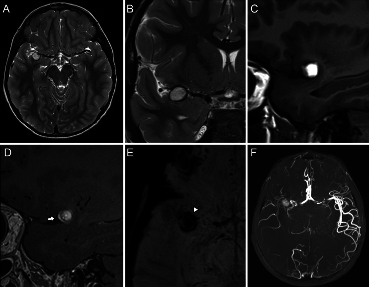
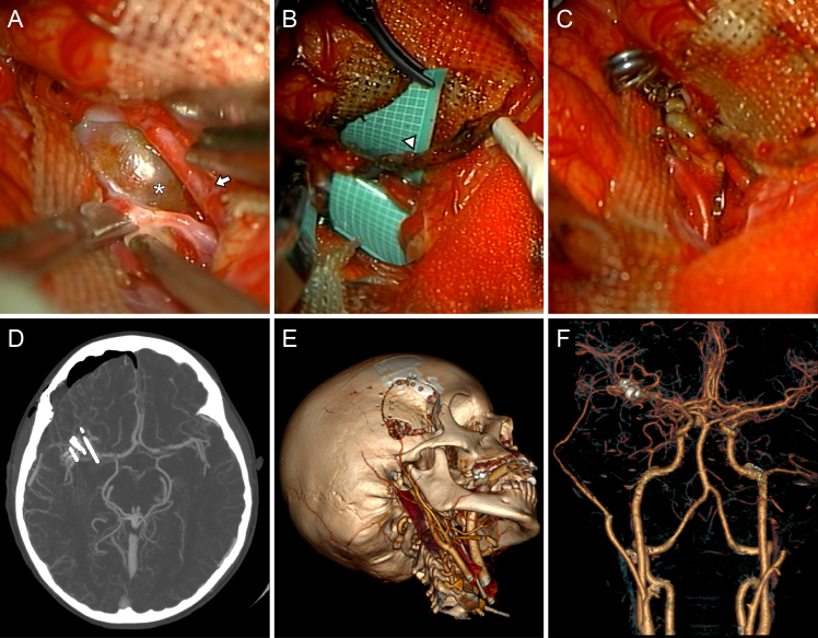
Pediatric intracranial aneurysms present unique diagnostic and therapeutic challenges due to their rarity and their distinct anatomical and physiological considerations compared with those of adult intracranial aneurysms. The authors present the case of a symptomatic pediatric patient who required emergency microsurgical treatment after a thrombosed dissecting aneurysm was identified in the right M1 segment of the middle cerebral artery.
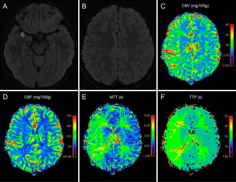
The lesion completely occluded its parent vessel, although distal blood flow was reconstituted through leptomeningeal collaterals. However, aneurysm wall contrast enhancement and signs of early perfusion changes were noticed, which prompted emergency treatment consisting of microsurgical aneurysm trapping, decompression, and extracranial/intracranial revascularization to be successfully performed through a minipterional craniotomy. After 1 year, the bypass occluded, although the patient remained asymptomatic. A slight enlargement of the ipsilateral anterior cerebral artery suggested the possibility of a benign hemodynamic rearrangement.
Emergency treatment may be necessary when signs of lesion instability or hemodynamic compromise are present; however, a comprehensive multidisciplinary evaluation is required. Treatment of complex vascular lesions using a minipterional approach is feasible even in pediatric patients, and delayed bypass occlusion may be a benign phenomenon reflecting gradual blood flow reorganization. https://thejns.org/doi/10.3171/CASE24484.
小儿颅内动脉瘤因其罕见性以及与成人颅内动脉瘤相比独特的解剖和生理因素,在诊断和治疗上面临着特殊挑战。作者报告了一例有症状的小儿患者病例,该患者在大脑中动脉右侧M1段发现一个血栓形成的夹层动脉瘤后,需要进行急诊显微手术治疗。
病变完全闭塞了其供血动脉,尽管通过软脑膜侧支循环重建了远端血流。然而,发现动脉瘤壁有对比增强以及早期灌注改变的迹象,这促使通过微创翼点入路成功实施了包括显微手术动脉瘤夹闭、减压以及颅外/颅内血管重建的急诊治疗。1年后,旁路闭塞,尽管患者仍无症状。同侧大脑前动脉略有增粗,提示可能存在良性血流动力学重新分布。
当出现病变不稳定或血流动力学受损的迹象时,可能需要进行急诊治疗;然而,需要进行全面的多学科评估。即使在小儿患者中,采用微创翼点入路治疗复杂血管病变也是可行的,旁路延迟闭塞可能是反映血流逐渐重新组织的良性现象。https://thejns.org/doi/10.3171/CASE24484