Department of Pharmaceutical Sciences, School of Pharmacy, College of Health and Human Sciences, North Dakota State University, Fargo, ND, 58102, USA.
Respir Res. 2024 Oct 28;25(1):387. doi: 10.1186/s12931-024-03017-4.
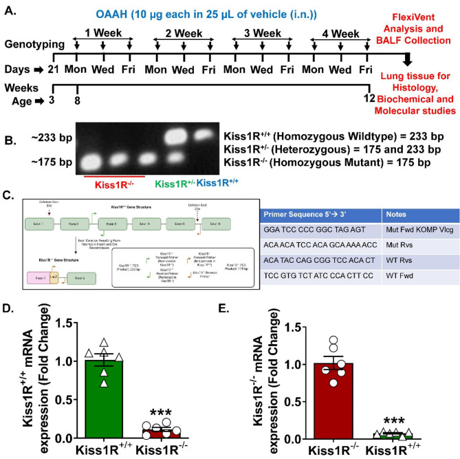
In asthma, sex-steroids signaling is recognized as a critical regulator of disease pathophysiology. However, the paradoxical role of sex-steroids, especially estrogen, suggests that an upstream mechanism or even independent of estrogen plays an important role in regulating asthma pathophysiology. In this context, in our previous studies, we explored kisspeptin (Kp) and its receptor Kiss1R's signaling in regulating human airway smooth muscle cell remodeling in vitro and airway hyperresponsiveness (AHR) in vivo in a mouse (wild-type, WT) model of asthma. In this study, we evaluated the effect of endogenous Kp in regulating AHR and remodeling using Kiss1R knockout (Kiss1R) mice.
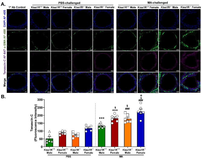
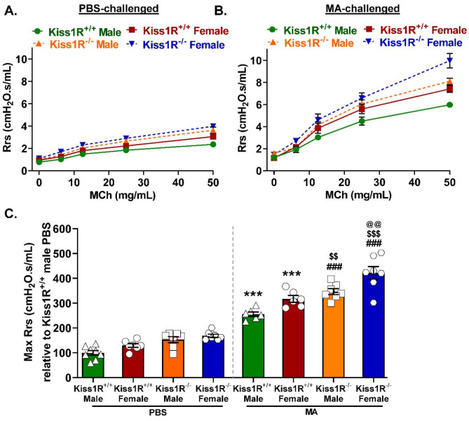
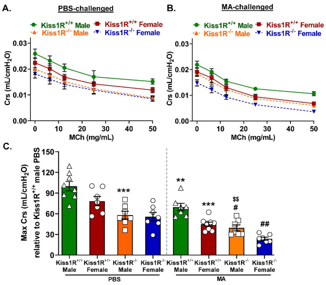
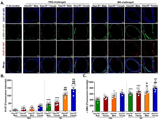
C57BL/6J WT (Kiss1R) and Kiss1R mice, both male and female, were intranasally challenged with mixed-allergen (MA) and/or phosphate-buffered saline (PBS). We used flexiVent analysis to assess airway resistance (Rrs), elastance (Ers), and compliance (Crs). Following this, broncho-alveolar lavage (BAL) was performed for differential leukocyte count (DLC) and cytokine analysis. Histology staining was performed using hematoxylin and eosin (H&E) for morphological analysis and Masson's Trichrome (MT) for collagen deposition. Additionally, lung sections were processed for immunofluorescence (IF) of Ki-67, α-smooth muscle actin (α-SMA), and tenascin-c.
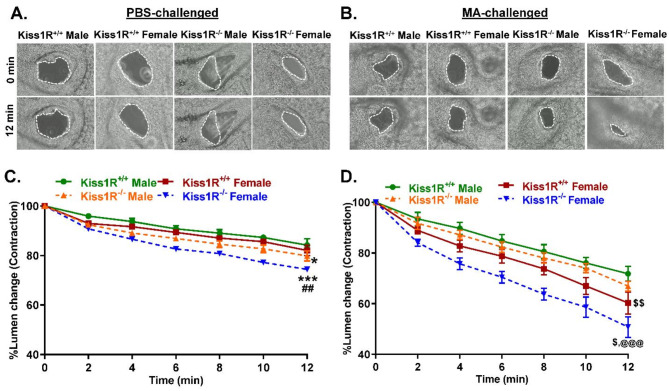
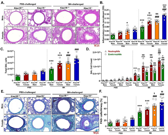
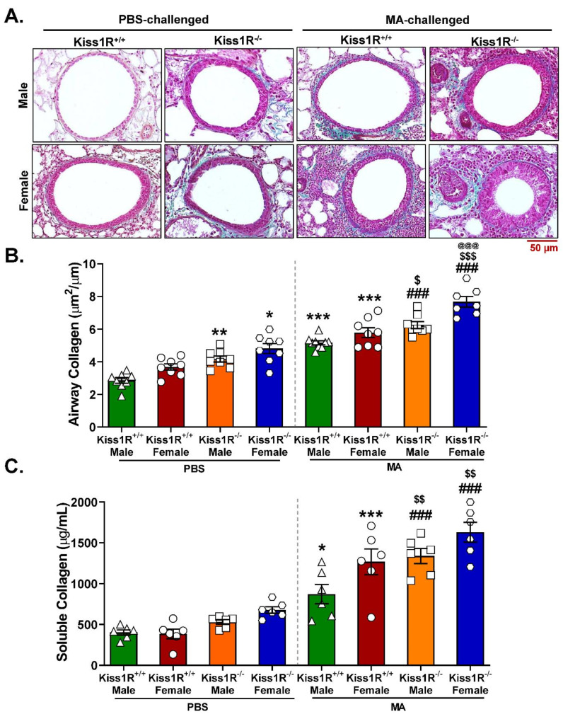
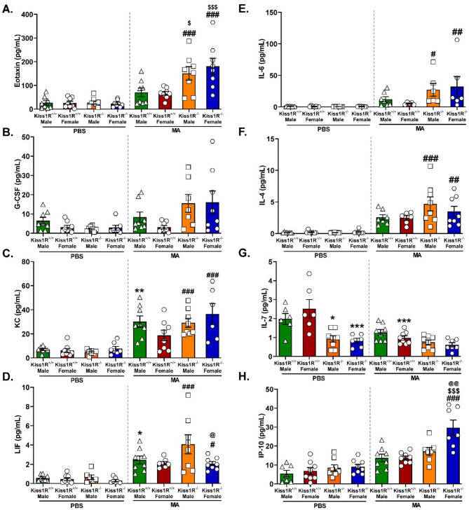
Interestingly, the loss of Kiss1R exacerbated lung function and airway contractility in mice challenged with MA, with more profound effects in Kiss1R female mice. MA-challenged Kiss1R mice showed a significant increase in immune cell infiltration and proinflammatory cytokine levels. Importantly, the loss of Kiss1R aggravated Th2/Th17 biased cytokines in MA-challenged mice. Furthermore, histology of lung sections from Kiss1R mice showed increased collagen deposition on airway walls and mucin production in airway cells compared to Kiss1R mice. In addition, immunofluorescence analysis showed loss of Kiss1R significantly aggravated airway remodeling and subsequently AHR.
These findings demonstrate the importance of inherent Kiss1R signaling in regulating airway inflammation, AHR, and remodeling in the pathophysiology of asthma.
在哮喘中,性激素信号被认为是疾病病理生理学的关键调节剂。然而,性激素的矛盾作用表明,上游机制甚至独立于雌激素在调节哮喘病理生理学中起着重要作用。在这方面,在我们之前的研究中,我们探索了 kisspeptin(Kp)及其受体 Kiss1R 在体外调节人气道平滑肌细胞重塑和体内调节哮喘小鼠模型气道高反应性(AHR)中的信号转导。在这项研究中,我们使用 Kiss1R 敲除(Kiss1R)小鼠评估内源性 Kp 调节 AHR 和重塑的作用。
雄性和雌性 C57BL/6J WT(Kiss1R)和 Kiss1R 小鼠均经鼻内给予混合过敏原(MA)和/或磷酸盐缓冲盐水(PBS)挑战。我们使用 flexiVent 分析评估气道阻力(Rrs)、弹性(Ers)和顺应性(Crs)。之后,进行支气管肺泡灌洗(BAL)以进行白细胞分类计数(DLC)和细胞因子分析。苏木精和伊红(H&E)染色用于形态学分析,马松三色(MT)染色用于胶原蛋白沉积。此外,对肺组织切片进行 Ki-67、α-平滑肌肌动蛋白(α-SMA)和 tenascin-c 的免疫荧光(IF)。
有趣的是,Kiss1R 的缺失加剧了 MA 挑战的小鼠的肺功能和气道收缩性,在 Kiss1R 雌性小鼠中影响更明显。MA 挑战的 Kiss1R 小鼠显示免疫细胞浸润和促炎细胞因子水平显著增加。重要的是,Kiss1R 的缺失加剧了 MA 挑战小鼠中的 Th2/Th17 偏向细胞因子。此外,Kiss1R 小鼠的肺组织切片的组织学显示气道壁上的胶原蛋白沉积和气道细胞中的粘蛋白产生增加。此外,免疫荧光分析表明,Kiss1R 的缺失显着加重了气道重塑,进而加重了 AHR。
这些发现表明固有 Kiss1R 信号在调节哮喘病理生理学中的气道炎症、AHR 和重塑中起着重要作用。