Kuroda Hidekatsu, Abe Tamami, Kamiyama Naohisa, Oguri Takuma, Ito Asami, Nakaya Ippeki, Watanabe Takuya, Abe Hiroaki, Yusa Kenji, Fujiwara Yudai, Sato Hiroki, Suzuki Akiko, Endo Kei, Yoshida Yuichi, Oikawa Takayoshi, Kakisaka Keisuke, Sawara Kei, Miyasaka Akio, Matsumoto Takayuki
Division of Gastroenterology and Hepatology, Department of Internal Medicine, Iwate Medical University, Iwate Medical University School of Medicine, Nishitokuta 2-1-1, Yahaba-Cho, Shiwa-Gun, Yahaba, Iwate, 028-3694, Japan.
Ultrasound General Imaging, GE HealthCare Japan, Hino-Shi, Japan.
J Gastroenterol. 2025 Feb;60(2):187-196. doi: 10.1007/s00535-024-02161-4. Epub 2024 Oct 29.
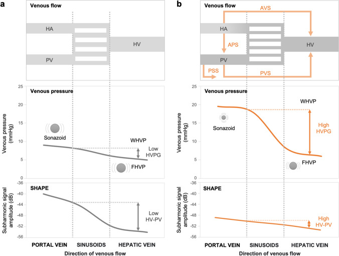
Subharmonic-aided pressure estimation (SHAPE) is a technique for determining changes in ambient pressure. We aimed to analyze a novel SHAPE integrated into ultrasound diagnostic equipment to predict patients with liver cirrhosis at high risk of esophagogastric varices (EV).
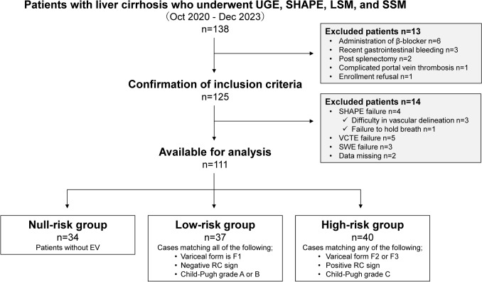
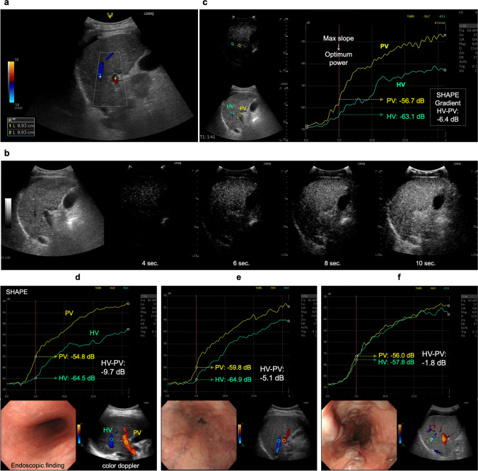
This prospective study included 111 patients with liver cirrhosis diagnosed between 2020 and 2023. We obtained liver stiffness measurements (LSM) and spleen stiffness measurements (SSM) using shear wave elastography and hepatic vein-portal vein (HV-PV) gradient using the SHAPE method. The EV risk was determined either as null, low, or high by esophagoscopy and Child-Pugh stage.
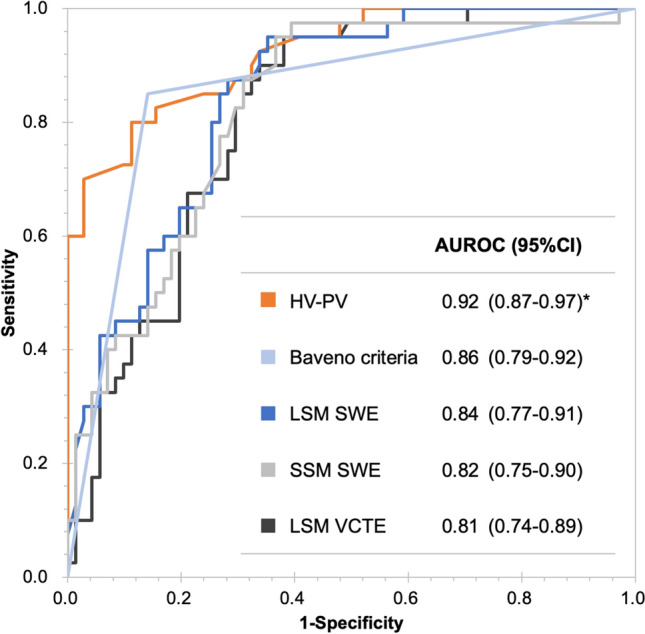
HV-PV gradient increased concordantly with the increase in EV risk (- 7.0 dB in null-risk, - 4.4 dB in low-risk, and - 2.0 dB in high-risk) with statistically significant difference among any two groups. The most appropriate cut-off value of the HV-PV gradient was - 3.5 dB, and sensitivity, specificity, and positive and negative predictive values were 80.0%, 89.0%, 80.0%, and 88.0%, respectively. The areas under the curve values for predicting the high-risk EV were 0.920, 0.843, and 0.824 for the HV-PV gradient, LSM, and SSM, respectively.
The novel SHAPE system demonstrated high accuracy in identifying patients with liver cirrhosis at a high risk of EV.
次谐波辅助压力估计(SHAPE)是一种用于确定环境压力变化的技术。我们旨在分析一种集成到超声诊断设备中的新型SHAPE,以预测患有食管胃静脉曲张(EV)高风险的肝硬化患者。
这项前瞻性研究纳入了2020年至2023年间诊断出的111例肝硬化患者。我们使用剪切波弹性成像获得肝脏硬度测量值(LSM)和脾脏硬度测量值(SSM),并使用SHAPE方法获得肝静脉-门静脉(HV-PV)梯度。通过食管镜检查和Child-Pugh分期将EV风险确定为无、低或高。
HV-PV梯度随着EV风险的增加而相应增加(无风险组为-7.0 dB,低风险组为-4.4 dB,高风险组为-2.0 dB),任意两组之间差异均有统计学意义。HV-PV梯度的最合适截断值为-3.5 dB,敏感性、特异性、阳性和阴性预测值分别为80.0%、89.0%、80.0%和88.0%。对于预测高风险EV,HV-PV梯度、LSM和SSM的曲线下面积值分别为0.920、0.843和0.824。
新型SHAPE系统在识别患有EV高风险的肝硬化患者方面显示出高准确性。