• 文献检索
  • 文档翻译
  • 深度研究
  • 学术资讯
  • Suppr Zotero 插件Zotero 插件
  • 邀请有礼
  • 套餐&价格
  • 历史记录
应用&插件
Suppr Zotero 插件Zotero 插件浏览器插件Mac 客户端Windows 客户端微信小程序
定价
高级版会员购买积分包购买API积分包
服务
文献检索文档翻译深度研究API 文档MCP 服务
关于我们
关于 Suppr公司介绍联系我们用户协议隐私条款
关注我们

Suppr 超能文献

核心技术专利:CN118964589B侵权必究
粤ICP备2023148730 号-1Suppr @ 2026

文献检索

告别复杂PubMed语法,用中文像聊天一样搜索,搜遍4000万医学文献。AI智能推荐,让科研检索更轻松。

立即免费搜索

文件翻译

保留排版,准确专业,支持PDF/Word/PPT等文件格式,支持 12+语言互译。

免费翻译文档

深度研究

AI帮你快速写综述,25分钟生成高质量综述,智能提取关键信息,辅助科研写作。

立即免费体验

[镓]Ga-NOTA-AE105 uPAR-PET/MRI在原发性胶质瘤患者中的前瞻性II期试验:预后价值及对uPAR靶向放射性核素治疗的意义

Prospective phase II trial of [Ga]Ga-NOTA-AE105 uPAR-PET/MRI in patients with primary gliomas: Prognostic value and Implications for uPAR-targeted Radionuclide Therapy.

作者信息

Azam Aleena, Kurbegovic Sorel, Carlsen Esben Andreas, Andersen Thomas Lund, Larsen Vibeke André, Law Ian, Skjøth-Rasmussen Jane, Kjaer Andreas

机构信息

Department of Clinical Physiology and Nuclear Medicine, Copenhagen University Hospital - Rigshospitalet, Blegdamsvej 9, Copenhagen, DK- 2100, Denmark.

Cluster for Molecular Imaging, Department of Biomedical Sciences, Copenhagen University Hospital - Rigshospitalet, University of Copenhagen, Copenhagen, Denmark.

出版信息

EJNMMI Res. 2024 Oct 29;14(1):100. doi: 10.1186/s13550-024-01164-9.

DOI:10.1186/s13550-024-01164-9
PMID:39472354
原文链接:https://pmc.ncbi.nlm.nih.gov/articles/PMC11522270/
Abstract

BACKGROUND

Treatment of patients with low-grade and high-grade gliomas is highly variable due to the large difference in survival expectancy. New non-invasive tools are needed for risk stratification prior to treatment. The urokinase plasminogen activator receptor (uPAR) is expressed in several cancers, associated with poor prognosis and may be non-invasively imaged using uPAR-PET. We aimed to investigate the uptake of the uPAR-PET tracer [Ga]Ga-NOTA-AE105 in primary gliomas and establish its prognostic value regarding overall survival (OS), and progression-free survival (PFS). Additionally, we analyzed the proportion of uPAR-PET positive tumors to estimate the potential number of candidates for future uPAR-PRRT.

METHODS

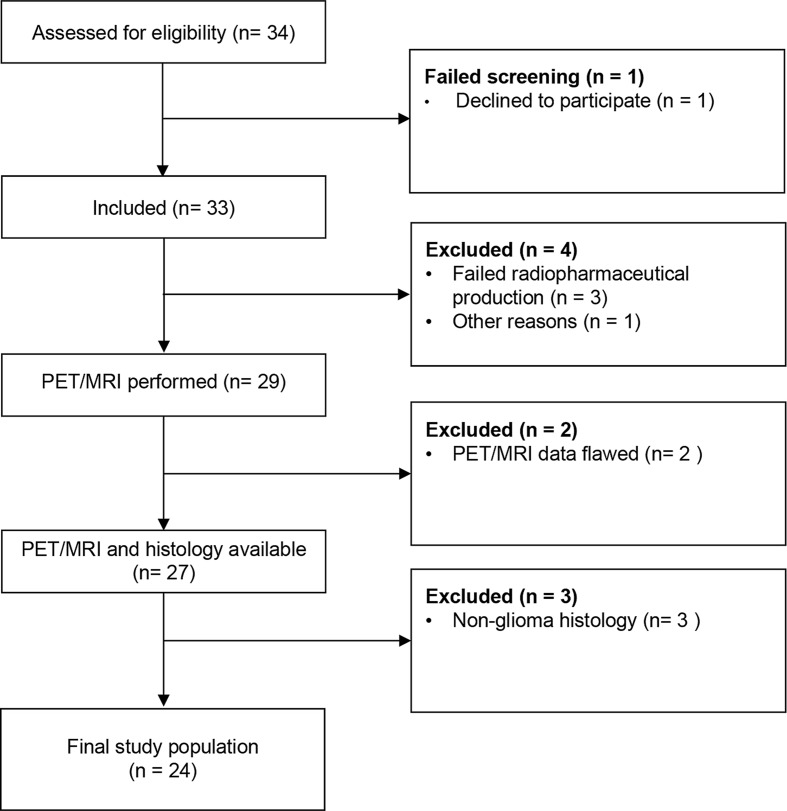
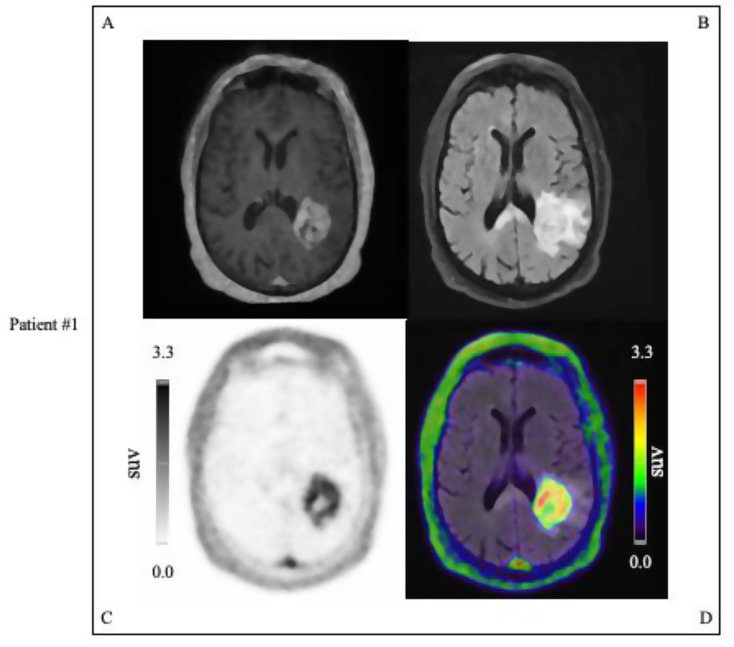
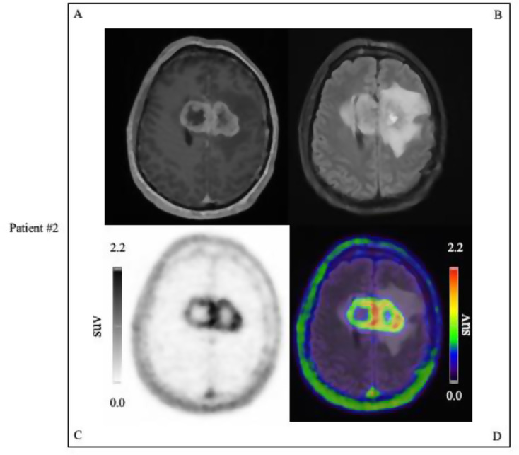
In a prospective phase II clinical trial, 24 patients suspected of primary glioma underwent a dynamic 60-min PET/MRI following the administration of approximately 200 MBq (range: 83-222 MBq) [Ga]Ga-NOTA-AE105. Lesions were considered uPAR positive if the tumor-to-background ratio, calculated as the ratio of TumorSUVmax-to-Normal-BrainSUVmean tumor-SUVmax-to-background-SUVmean, was ≥ 2.0. The patients were followed over time to assess OS and PFS and stratified into high and low uPAR expression groups based on TumorSUVmax.

RESULTS

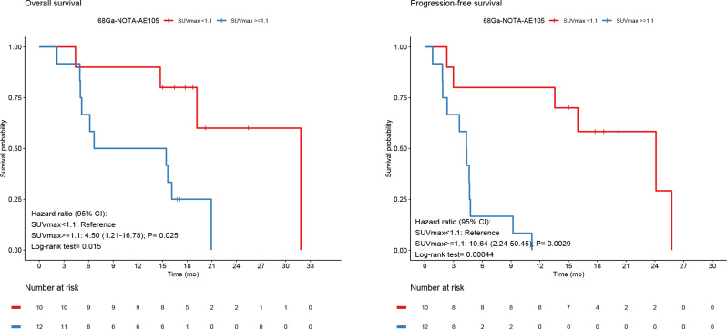
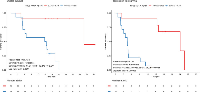
Of the 24 patients, 16 (67%) were diagnosed with WHO grade 4 gliomas, 6 (25%) with grade 3, and 2 (8%) with grade 2. Two-thirds of all patients (67%) presented with uPAR positive lesions and 94% grade 4 gliomas. At median follow up of 18.8 (2.1-45.6) months, 19 patients had disease progression and 14 had died. uPAR expression dichotomized into high and low, revealed significant worse prognosis for the high uPAR group for OS and PFS with HR of 14.3 (95% CI, 1.8-112.3; P = 0.011), and HR of 26.5 (95% CI, 3.3-214.0; P = 0.0021), respectively. uPAR expression as a continuous variable was associated with worse prognosis for OS and PFS with HR of 2.7 (95% CI, 1.5-4.8; P = 0.0012), and HR of 2.5 (95% CI, 1.5-4.2; P = 0.00073), respectively.

CONCLUSIONS

The majority of glioma patients and almost all with grade 4 gliomas displayed uPAR positive lesions underlining the feasibility of Ga-NOTA-AE105 PET/MRI in gliomas. High uPAR expression is significantly correlated with worse survival outcomes for patients. Additionally, the high proportion of uPAR positive gliomas underscores the potential of uPAR-targeted radionuclide therapy in these patients.

TRAIL REGISTRATION

EudraCT No: 2016-002417-21; the Scientific Ethics Committee: H-16,035,303; the Danish Data Protection Agency: 2012-58-0004; clinical trials registry: NCT02945826, 26Oct2016, URL: https://classic.

CLINICALTRIALS

gov/ct2/show/NCT02945826 .

摘要

背景

由于低级别和高级别胶质瘤患者的生存预期差异很大,其治疗方式高度可变。在治疗前需要新的非侵入性工具进行风险分层。尿激酶型纤溶酶原激活物受体(uPAR)在多种癌症中表达,与预后不良相关,并且可以使用uPAR-PET进行非侵入性成像。我们旨在研究uPAR-PET示踪剂[镓]Ga-NOTA-AE105在原发性胶质瘤中的摄取情况,并确定其对总生存期(OS)和无进展生存期(PFS)的预后价值。此外,我们分析了uPAR-PET阳性肿瘤的比例,以估计未来uPAR-PRRT候选者的潜在数量。

方法

在一项前瞻性II期临床试验中,24例疑似原发性胶质瘤患者在静脉注射约200MBq(范围:83-222MBq)[镓]Ga-NOTA-AE105后接受了60分钟的动态PET/MRI检查。如果肿瘤与背景比值(计算为肿瘤SUVmax与正常脑SUVmean之比,即肿瘤SUVmax与背景SUVmean之比)≥2.0,则病变被认为是uPAR阳性。对患者进行随访以评估OS和PFS,并根据肿瘤SUVmax将其分为uPAR高表达组和低表达组。

结果

24例患者中,16例(67%)被诊断为WHO 4级胶质瘤,6例(25%)为3级,2例(8%)为2级。所有患者中有三分之二(67%)出现uPAR阳性病变,4级胶质瘤患者中这一比例为94%。在中位随访18.8(2.1-45.6)个月时,19例患者出现疾病进展,14例死亡。将uPAR表达分为高表达和低表达,结果显示高uPAR组的OS和PFS预后明显更差,HR分别为14.3(95%CI,1.8-112.3;P=0.011)和26.5(95%CI,3.3-214.0;P=0.0021)。uPAR表达作为连续变量与OS和PFS的预后较差相关,HR分别为2.7(95%CI,1.5-4.8;P=0.0012)和2.5(95%CI,1.5-4.2;P=0.00073)。

结论

大多数胶质瘤患者以及几乎所有4级胶质瘤患者均表现为uPAR阳性病变,这突出了Ga-NOTA-AE105 PET/MRI在胶质瘤中的可行性。高uPAR表达与患者较差的生存结果显著相关。此外,高比例的uPAR阳性胶质瘤强调了uPAR靶向放射性核素治疗在这些患者中的潜力。

试验注册

EudraCT编号:2016-002417-21;科学伦理委员会:H-16,035,303;丹麦数据保护局:2012-58-0004;临床试验注册:NCT02945826,2016年10月26日,网址:https://classic.

CLINICALTRIALS

gov/ct2/show/NCT02945826 。

https://cdn.ncbi.nlm.nih.gov/pmc/blobs/e034/11522270/e960d7ca8c42/13550_2024_1164_Fig7_HTML.jpg
https://cdn.ncbi.nlm.nih.gov/pmc/blobs/e034/11522270/2086a3025da8/13550_2024_1164_Fig1_HTML.jpg
https://cdn.ncbi.nlm.nih.gov/pmc/blobs/e034/11522270/14a713c8e6f2/13550_2024_1164_Fig2_HTML.jpg
https://cdn.ncbi.nlm.nih.gov/pmc/blobs/e034/11522270/597c9446f8d6/13550_2024_1164_Fig3_HTML.jpg
https://cdn.ncbi.nlm.nih.gov/pmc/blobs/e034/11522270/8b580fd0b8a5/13550_2024_1164_Fig4_HTML.jpg
https://cdn.ncbi.nlm.nih.gov/pmc/blobs/e034/11522270/0e18a6bcfb4e/13550_2024_1164_Fig5_HTML.jpg
https://cdn.ncbi.nlm.nih.gov/pmc/blobs/e034/11522270/02abb26f133e/13550_2024_1164_Fig6_HTML.jpg
https://cdn.ncbi.nlm.nih.gov/pmc/blobs/e034/11522270/e960d7ca8c42/13550_2024_1164_Fig7_HTML.jpg

相似文献

1
Prospective phase II trial of [Ga]Ga-NOTA-AE105 uPAR-PET/MRI in patients with primary gliomas: Prognostic value and Implications for uPAR-targeted Radionuclide Therapy.[镓]Ga-NOTA-AE105 uPAR-PET/MRI在原发性胶质瘤患者中的前瞻性II期试验:预后价值及对uPAR靶向放射性核素治疗的意义
EJNMMI Res. 2024 Oct 29;14(1):100. doi: 10.1186/s13550-024-01164-9.
2
Prospective Phase II Trial of Prognostication by Ga-NOTA-AE105 uPAR PET in Patients with Neuroendocrine Neoplasms: Implications for uPAR-Targeted Therapy.神经内分泌肿瘤患者 Ga-NOTA-AE105 uPAR PET 预后预测的前瞻性 II 期试验:对 uPAR 靶向治疗的影响。
J Nucl Med. 2022 Sep;63(9):1371-1377. doi: 10.2967/jnumed.121.263177. Epub 2022 Jan 20.
3
Prognostic Value of Urokinase-Type Plasminogen Activator Receptor PET/CT in Head and Neck Squamous Cell Carcinomas and Comparison with F-FDG PET/CT: A Single-Center Prospective Study.尿激酶型纤溶酶原激活物受体 PET/CT 对头颈部鳞状细胞癌的预后价值及与 F-FDG PET/CT 的比较:一项单中心前瞻性研究。
J Nucl Med. 2022 Aug;63(8):1169-1176. doi: 10.2967/jnumed.121.262866. Epub 2021 Dec 2.
4
Urokinase-Type Plasminogen Activator Receptor as a Potential PET Biomarker in Glioblastoma.尿激酶型纤溶酶原激活物受体作为胶质母细胞瘤潜在的正电子发射断层显像生物标志物
J Nucl Med. 2016 Feb;57(2):272-8. doi: 10.2967/jnumed.115.161703. Epub 2015 Oct 1.
5
Diagnostic Value of Preoperative uPAR-PET/CT in Regional Lymph Node Staging of Oral and Oropharyngeal Squamous Cell Carcinoma: A Prospective Phase II Trial.术前uPAR-PET/CT在口腔和口咽鳞状细胞癌区域淋巴结分期中的诊断价值:一项前瞻性II期试验
Diagnostics (Basel). 2023 Oct 25;13(21):3303. doi: 10.3390/diagnostics13213303.
6
Safety, Dosimetry, and Tumor Detection Ability of Ga-NOTA-AE105: First-in-Human Study of a Novel Radioligand for uPAR PET Imaging.Ga-NOTA-AE105的安全性、剂量学及肿瘤检测能力:一种用于uPAR正电子发射断层显像(PET)成像的新型放射性配体的首次人体研究。
J Nucl Med. 2017 Mar;58(3):379-386. doi: 10.2967/jnumed.116.178970. Epub 2016 Sep 8.
7
68Ga-labeling and in vivo evaluation of a uPAR binding DOTA- and NODAGA-conjugated peptide for PET imaging of invasive cancers.68Ga 标记及体内评价一种用于侵袭性癌症 PET 成像的 uPAR 结合的 DOTA 和 NODAGA 缀合肽。
Nucl Med Biol. 2012 May;39(4):560-9. doi: 10.1016/j.nucmedbio.2011.10.011. Epub 2011 Dec 14.
8
First (18)F-labeled ligand for PET imaging of uPAR: in vivo studies in human prostate cancer xenografts.首个用于 uPAR 的正电子发射断层扫描(PET)成像的 18F 标记配体:在人前列腺癌异种移植模型中的体内研究。
Nucl Med Biol. 2013 Jul;40(5):618-24. doi: 10.1016/j.nucmedbio.2013.03.001. Epub 2013 Apr 18.
9
68 Ga-PSMA-11 PET/CT Imaging in Brain Gliomas and Its Correlation With Clinicopathological Prognostic Parameters.68Ga-PSMA-11 PET/CT 成像在脑胶质瘤中的应用及其与临床病理预后参数的相关性。
Clin Nucl Med. 2023 Dec 1;48(12):e559-e563. doi: 10.1097/RLU.0000000000004903. Epub 2023 Oct 23.
10
uPAR PET/CT for Prognostication and Response Assessment in Patients with Metastatic Castration-Resistant Prostate Cancer Undergoing Radium-223 Therapy: A Prospective Phase II Study.用于接受镭-223治疗的转移性去势抵抗性前列腺癌患者预后评估和疗效评估的uPAR正电子发射断层扫描/计算机断层扫描:一项前瞻性II期研究
Diagnostics (Basel). 2021 Jun 14;11(6):1087. doi: 10.3390/diagnostics11061087.

引用本文的文献

1
The molecular blueprint of targeted radionuclide therapy.靶向放射性核素治疗的分子蓝图。
Nat Rev Clin Oncol. 2025 Sep 9. doi: 10.1038/s41571-025-01069-z.
2
Trigeminal nerve-driven neurogenic inflammation linking migraine to glioblastoma invasion: a literature review.三叉神经驱动的神经源性炎症将偏头痛与胶质母细胞瘤侵袭联系起来:文献综述
Front Immunol. 2025 Jul 16;16:1632154. doi: 10.3389/fimmu.2025.1632154. eCollection 2025.

本文引用的文献

1
Diagnostic Value of Preoperative uPAR-PET/CT in Regional Lymph Node Staging of Oral and Oropharyngeal Squamous Cell Carcinoma: A Prospective Phase II Trial.术前uPAR-PET/CT在口腔和口咽鳞状细胞癌区域淋巴结分期中的诊断价值:一项前瞻性II期试验
Diagnostics (Basel). 2023 Oct 25;13(21):3303. doi: 10.3390/diagnostics13213303.
2
Prospective Phase II Trial of Prognostication by Ga-NOTA-AE105 uPAR PET in Patients with Neuroendocrine Neoplasms: Implications for uPAR-Targeted Therapy.神经内分泌肿瘤患者 Ga-NOTA-AE105 uPAR PET 预后预测的前瞻性 II 期试验:对 uPAR 靶向治疗的影响。
J Nucl Med. 2022 Sep;63(9):1371-1377. doi: 10.2967/jnumed.121.263177. Epub 2022 Jan 20.
3
Prognostic Value of Urokinase-Type Plasminogen Activator Receptor PET/CT in Head and Neck Squamous Cell Carcinomas and Comparison with F-FDG PET/CT: A Single-Center Prospective Study.
尿激酶型纤溶酶原激活物受体 PET/CT 对头颈部鳞状细胞癌的预后价值及与 F-FDG PET/CT 的比较:一项单中心前瞻性研究。
J Nucl Med. 2022 Aug;63(8):1169-1176. doi: 10.2967/jnumed.121.262866. Epub 2021 Dec 2.
4
Peptide Receptor Radionuclide Therapy and Primary Brain Tumors: An Overview.肽受体放射性核素治疗与原发性脑肿瘤:概述
Pharmaceuticals (Basel). 2021 Aug 29;14(9):872. doi: 10.3390/ph14090872.
5
The 2021 WHO Classification of Tumors of the Central Nervous System: a summary.2021 年世卫组织中枢神经系统肿瘤分类:概述。
Neuro Oncol. 2021 Aug 2;23(8):1231-1251. doi: 10.1093/neuonc/noab106.
6
IRDye800CW labeled uPAR-targeting peptide for fluorescence-guided glioblastoma surgery: Preclinical studies in orthotopic xenografts.IRDye800CW标记的靶向尿激酶型纤溶酶原激活物受体(uPAR)的肽用于荧光引导的胶质母细胞瘤手术:原位异种移植的临床前研究
Theranostics. 2021 May 21;11(15):7159-7174. doi: 10.7150/thno.49787. eCollection 2021.
7
Fibrinolytic System and Cancer: Diagnostic and Therapeutic Applications.纤维蛋白溶解系统与癌症:诊断与治疗应用。
Int J Mol Sci. 2021 Apr 22;22(9):4358. doi: 10.3390/ijms22094358.
8
EANO guidelines on the diagnosis and treatment of diffuse gliomas of adulthood.EANO 成人弥漫性胶质瘤诊断与治疗指南。
Nat Rev Clin Oncol. 2021 Mar;18(3):170-186. doi: 10.1038/s41571-020-00447-z. Epub 2020 Dec 8.
9
Urokinase-Type Plasminogen Activator Receptor (uPAR) PET/MRI of Prostate Cancer for Noninvasive Evaluation of Aggressiveness: Comparison with Gleason Score in a Prospective Phase 2 Clinical Trial.尿激酶型纤溶酶原激活物受体(uPAR)前列腺癌 PET/MRI 非侵入性评估侵袭性:与前瞻性 2 期临床试验中 Gleason 评分的比较。
J Nucl Med. 2021 Mar;62(3):354-359. doi: 10.2967/jnumed.120.248120. Epub 2020 Aug 6.
10
AI-driven attenuation correction for brain PET/MRI: Clinical evaluation of a dementia cohort and importance of the training group size.人工智能驱动的脑 PET/MRI 衰减校正:痴呆队列的临床评估及训练组规模的重要性。
Neuroimage. 2020 Nov 15;222:117221. doi: 10.1016/j.neuroimage.2020.117221. Epub 2020 Aug 1.