Department of Urinary and Vascular Imaging, Hôpital Edouard Herriot, Hospices Civils de Lyon, Lyon, France.
Medisys, Philips Research Paris, Paris, France.
Eur Radiol Exp. 2024 Oct 30;8(1):122. doi: 10.1186/s41747-024-00520-7.
Our aim was to train and test a deep learning-based algorithm for automatically segmenting kidneys and renal cysts in patients with autosomal dominant polycystic kidney disease (ADPKD).
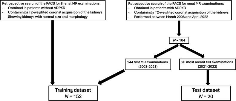
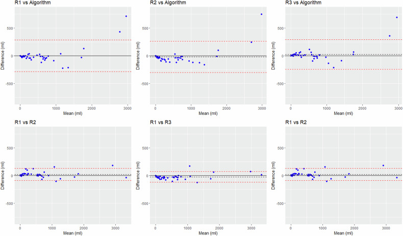
We retrospectively selected all ADPKD patients who underwent renal MRI with coronal T2-weighted imaging at our institution from 2008 to 2022. The 20 most recent examinations constituted the test dataset, to mimic pseudoprospective enrolment. The remaining ones constituted the training dataset to which eight normal renal MRIs were added. Kidneys and cysts ground truth segmentations were performed on coronal T2-weighted images by a junior radiologist supervised by an experienced radiologist. Kidneys and cysts of the 20 test MRIs were segmented by the algorithm and three independent human raters. Segmentations were compared using overlap metrics. The total kidney volume (TKV), total cystic volume (TCV), and cystic index (TCV divided by TKV) were compared using Bland-Altman analysis.
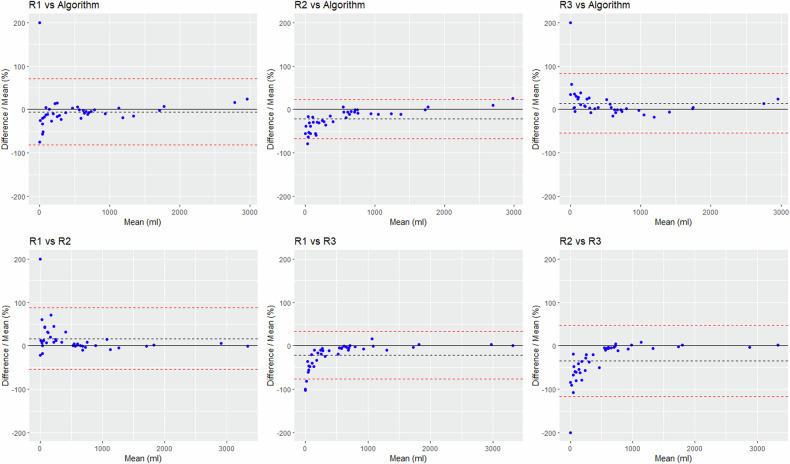
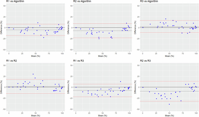
We included 164 ADPKD patients. Dice similarity coefficients ranged from 85.9% to 87.4% between the algorithms and the raters' segmentations and from 84.2% to 86.2% across raters' segmentations. For TCV assessment, the biases ± standard deviations (SD) were 3-19 ± 137-151 mL between the algorithm and the raters, and 22-45 ± 49-57 mL across raters. The algorithm underestimated TKV and TCV in two outliers with TCV > 2800 mL. For cystic index assessment, the biases ± SD were 2.5-6.9% ± 6.7-8.3% between the algorithm and the raters, and 2.1-9.4 ± 7.4-11.6% across raters.
The algorithm's performance fell within the range of inter-rater variability, but large TKV and TCV were underestimated.
Accurate automated segmentation of the renal cysts will enable the large-scale evaluation of the prognostic value of TCV and cystic index in ADPKD patients. If these biomarkers are prognostic, then automated segmentation will facilitate their use in daily routine.
Cystic volume is an emerging biomarker in ADPKD. The algorithm's performance in segmenting kidneys and cysts fell within interrater variability. The segmentation of very large cysts, under-represented in the training dataset, needs improvement.
我们的目的是训练和测试一种基于深度学习的算法,以自动分割常染色体显性多囊肾病(ADPKD)患者的肾脏和肾囊肿。
我们回顾性地选择了 2008 年至 2022 年期间在我院接受冠状 T2 加权 MRI 的所有 ADPKD 患者。最近的 20 次检查构成了测试数据集,以模拟前瞻性入组。其余的构成了训练数据集,并添加了 8 个正常的肾脏 MRI。初级放射科医师在有经验的放射科医师的指导下对冠状 T2 加权图像上的肾脏和囊肿进行了ground truth 分割。对 20 个测试 MRI 的肾脏和囊肿进行了算法和三个独立的人工评分者的分割。使用重叠度量来比较分割。使用 Bland-Altman 分析比较总肾体积(TKV)、总囊肿体积(TCV)和囊肿指数(TCV 除以 TKV)。
我们纳入了 164 名 ADPKD 患者。算法与评分者分割之间的 Dice 相似系数范围为 85.9%至 87.4%,评分者之间的分割范围为 84.2%至 86.2%。对于 TCV 评估,算法与评分者之间的偏差±标准偏差(SD)为 3-19±137-151mL,评分者之间的偏差为 22-45±49-57mL。算法低估了两个 TCV>2800mL 的异常值的 TKV 和 TCV。对于囊肿指数评估,算法与评分者之间的偏差±SD 为 2.5-6.9%±6.7-8.3%,评分者之间的偏差为 2.1-9.4%±7.4-11.6%。
算法的性能在评分者之间的可变性范围内,但对大 TKV 和 TCV 的估计偏低。
对肾囊肿的自动分割将能够大规模评估 TCV 和囊肿指数在 ADPKD 患者中的预后价值。如果这些生物标志物具有预后意义,那么自动分割将有助于在日常工作中使用它们。
囊肿体积是 ADPKD 的一个新兴生物标志物。算法在分割肾脏和囊肿方面的性能在评分者之间的可变性范围内。需要改进对训练数据集中代表性不足的非常大的囊肿的分割。