Khandelwal Shreya, Dhande Rajasbala, Parihar Pratapsingh, Mishra Gaurav V, Sood Anshul
Radiodiagnosis, Jawaharlal Nehru Medical College, Datta Meghe Institute of Higher Education and Research (DMIHER), Wardha, IND.
Cureus. 2024 Oct 1;16(10):e70596. doi: 10.7759/cureus.70596. eCollection 2024 Oct.
This study aims to evaluate the role of multidetector computed tomography (MDCT) urography in cases of obstructive uropathy to determine the cause, side, site, and level of obstruction and to differentiate between acute and chronic cases of obstructive uropathy based on imaging features.
Using Cochran's formula, a sample size of 121 patients was calculated. The patients underwent computed tomography (CT) urography to assess the obstructing agents causing obstructive uropathy. The conducted scan had four phases: the non-contrast phase, corticomedullary phase, nephrographic phase, and excretory phase. We assessed the obstructive agents and the changes they caused in the urinary tract.
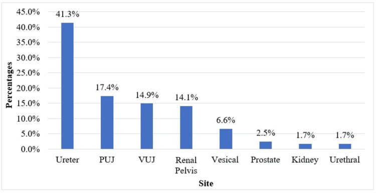
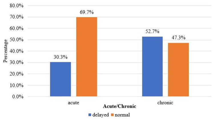
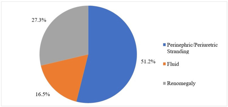
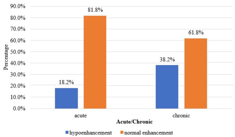
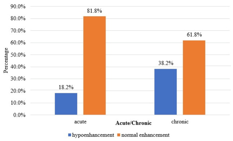
A total of 74 patients (61.16%) had calculus as their obstructive agent, followed by stricture (14.88%). The obstructive agents were intraluminal in 102 patients (84.3%) and extraluminal in 19 patients (15.7%). The ureter was the most common site of obstruction, accounting for 41.32%. The acute cases were 66 (54.55%), and the chronic cases were 55 (45.45%). A statistically significant (p<0.05) association was found using the chi-square test in the comparison of the enhancement and excretion of the kidneys and the type of case (acute or chronic). A statistically significant (p<0.05) association was found using the chi-square test in the comparison of the distribution of the secondary findings, such as perinephric fat stranding and perinephric fluid collection, and the type of case (acute or chronic).
MDCT urography is a highly reliable method of imaging the cause of obstructing agents in cases of obstructive uropathy and the damage caused by them. The type of enhancement and excretion and the secondary findings play an important role in determining the acuteness or the chronicity of the obstructive agent.
本研究旨在评估多排螺旋计算机断层扫描(MDCT)尿路造影在梗阻性肾病病例中的作用,以确定梗阻的原因、侧别、部位和程度,并根据影像学特征区分梗阻性肾病的急性和慢性病例。
使用 Cochr an公式计算出样本量为121例患者。患者接受计算机断层扫描(CT)尿路造影以评估导致梗阻性肾病的梗阻因素。所进行的扫描有四个阶段:非增强期、皮质髓质期、肾实质期和排泄期。我们评估了梗阻因素及其在尿路中引起的变化。
共有74例患者(61.16%)的梗阻因素为结石,其次是狭窄(14.88%)。102例患者(84.3%)的梗阻因素位于管腔内,19例患者(15.7%)位于管腔外。输尿管是最常见的梗阻部位,占41.32%。急性病例有66例(54.55%),慢性病例有55例(45.45%)。在比较肾脏的强化和排泄情况与病例类型(急性或慢性)时,使用卡方检验发现有统计学意义(p<0.05)的关联。在比较诸如肾周脂肪条索和肾周液体积聚等次要表现的分布情况与病例类型(急性或慢性)时,使用卡方检验发现有统计学意义(p<0.05)的关联。
MDCT尿路造影是一种高度可靠的成像方法,可用于显示梗阻性肾病病例中梗阻因素的原因及其所造成的损害。强化和排泄的类型以及次要表现对于确定梗阻因素的急性或慢性程度起着重要作用。