Garg Anjali, Vo Sheeny, Brase Logan, Aladyeva Ekaterina, Albanus Ricardo D'O, Nallapu Aasritha, Fu Hongjun, Harari Oscar
Department of Psychiatry, Washington University, Saint Louis, St. Louis, Missouri, United States of America.
Department of Neuroscience, OSU Wexner Medical Center, The Ohio State University, Columbus, OH, United States of America.
Res Sq. 2024 Oct 16:rs.3.rs-5045715. doi: 10.21203/rs.3.rs-5045715/v1.
Substantial evidence has established the critical role of microglia, the brain's resident immune cells, in the pathogenesis of Alzheimer's disease (AD). Microglia exhibit diverse transcriptional states in response to neuroinflammatory stimuli, and understanding these states is crucial for elucidating the underlying mechanisms of AD.
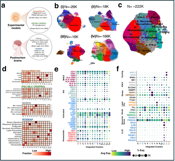
In this work, we integrated single-cell and spatially resolved transcriptomics data from multiple cohorts and brain regions, including microglia from experimental and human brains.
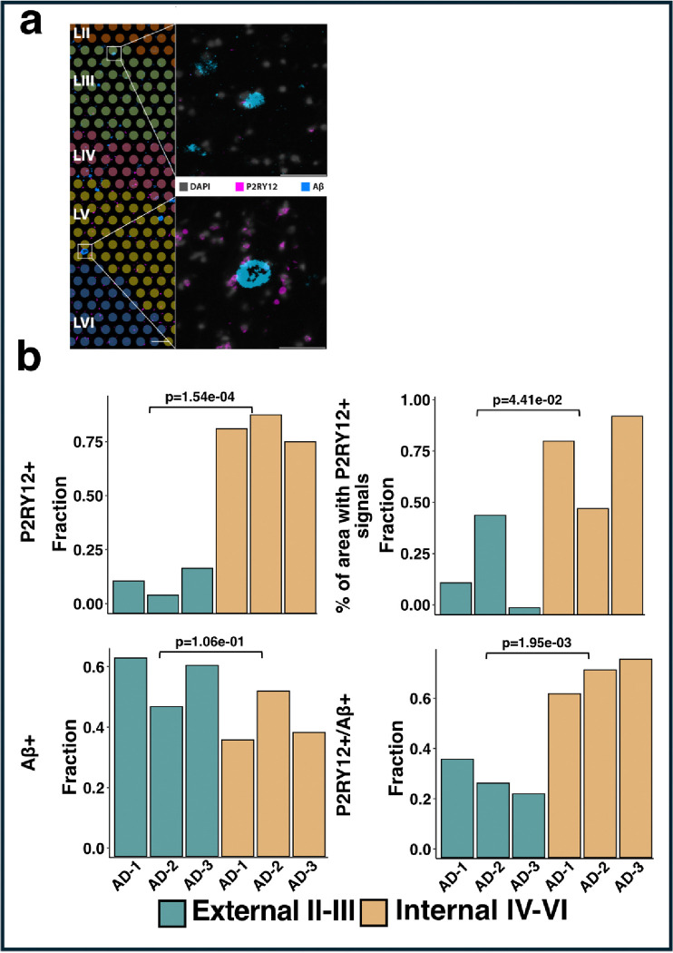
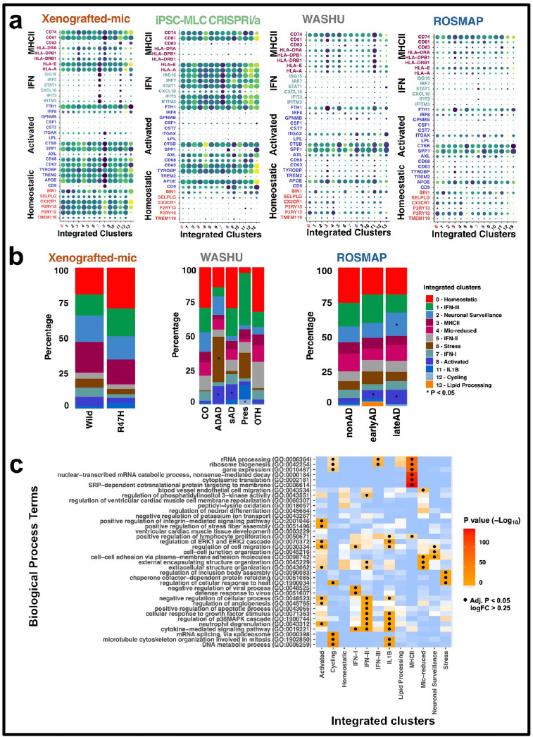
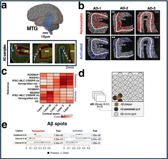
This comprehensive atlas revealed a great heterogeneity of microglial states, with a significant enrichment of specific states, including activated microglia, in AD brains compared to controls. Further integration of spatial transcriptomics and immunohistochemistry showed that activated microglia are predominantly located in the external cortical layers near amyloid plaques, while homeostatic microglia are more prevalent in the internal cortical layers and further away from the plaques. These spatial patterns were further validated using P2RY12 immunostaining, which confirmed the reliability of the transcriptomic data.
By integrating single-cell and spatial transcriptomics, we have provided a detailed atlas of microglial diversity, revealing the regional and pathological specificity of microglial states.
大量证据表明,作为大脑常驻免疫细胞的小胶质细胞在阿尔茨海默病(AD)的发病机制中起关键作用。小胶质细胞会对神经炎症刺激表现出多种转录状态,了解这些状态对于阐明AD的潜在机制至关重要。
在这项研究中,我们整合了来自多个队列和脑区的单细胞及空间分辨转录组学数据,包括来自实验性和人类大脑的小胶质细胞。
这个综合图谱揭示了小胶质细胞状态的高度异质性,与对照组相比,AD大脑中特定状态(包括活化的小胶质细胞)显著富集。空间转录组学和免疫组织化学的进一步整合表明,活化的小胶质细胞主要位于淀粉样斑块附近的皮质外层,而稳态小胶质细胞在皮质内层更为普遍且离斑块更远。这些空间模式通过P2RY12免疫染色得到进一步验证,证实了转录组数据的可靠性。
通过整合单细胞和空间转录组学,我们提供了一份详细的小胶质细胞多样性图谱,揭示了小胶质细胞状态的区域和病理特异性。