Department of Molecular Biosciences, Davis, School of Veterinary Medicine, University of California, Davis, CA, 95616, USA.
Center for Molecular and Genomic Imaging, College of Engineering, University of California, DavisDavis, CA, 95616, USA.
J Neuroinflammation. 2024 Nov 4;21(1):285. doi: 10.1186/s12974-024-03272-8.
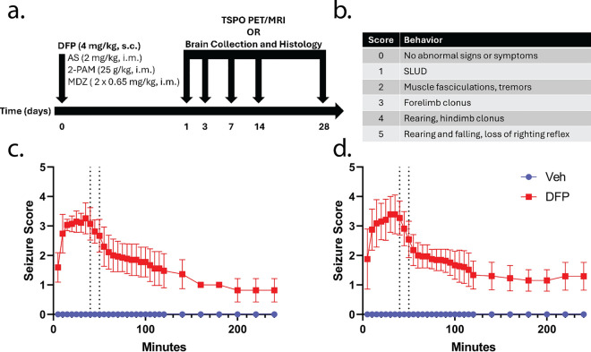
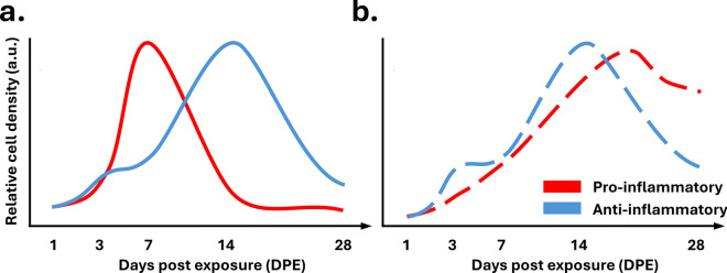
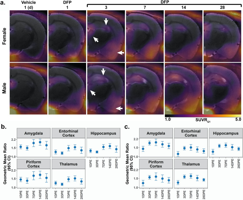
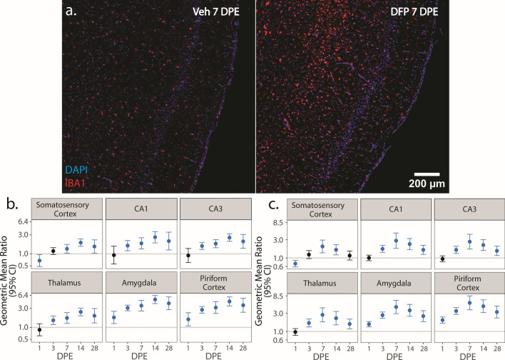
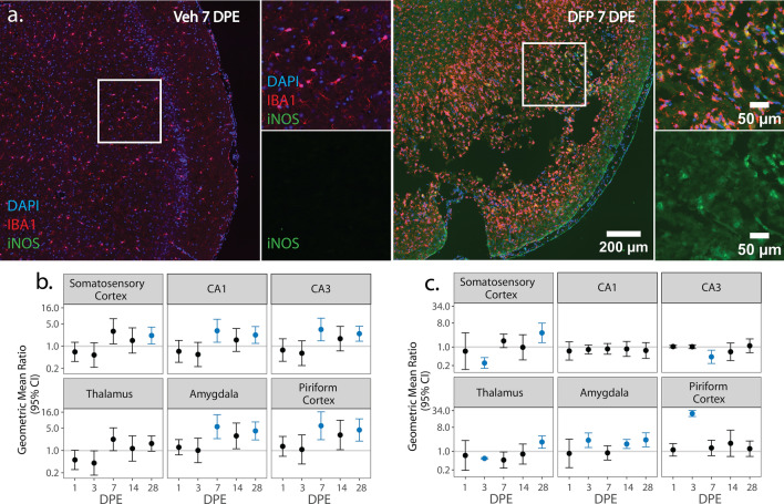
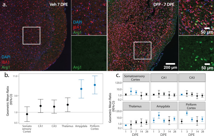
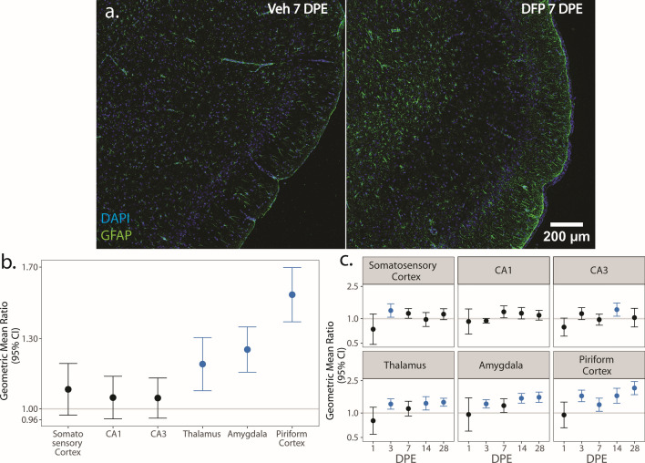
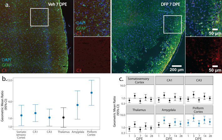
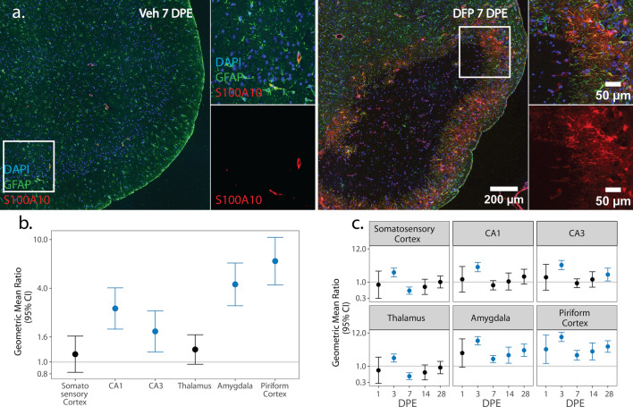
Acute intoxication with cholinesterase inhibiting organophosphates (OP) can produce life-threatening cholinergic crisis and status epilepticus (SE). Survivors often develop long-term neurological consequences, including spontaneous recurrent seizures (SRS) and impaired cognition. Numerous studies implicate OP-induced neuroinflammation as a pathogenic mechanism contributing to these chronic sequelae; however, little is known about the inflammatory phenotype of innate immune cells in the brain following acute OP intoxication. Thus, the aim of this study was to characterize the natural history of microglial and astrocytic inflammatory phenotypes following acute intoxication with the OP, diisopropylfluorophosphate (DFP). Adult male and female Sprague-Dawley rats were administered a single dose of DFP (4 mg/kg, sc) followed by standard medical countermeasures. Within minutes, animals developed benzodiazepine-resistant SE as determined by monitoring seizures using a modified Racine scale. At 1, 3, 7, 14, and 28 d post-exposure (DPE), neuroinflammation was assessed using translocator protein (TSPO) positron emission tomography (PET) and magnetic resonance imaging (MRI). In both sexes, we observed consistently elevated radiotracer uptake across all examined brain regions and time points. A separate group of animals was euthanized at these same time points to collect tissues for immunohistochemical analyses. Colocalization of IBA-1, a marker for microglia, with iNOS or Arg1 was used to identify pro- and anti-inflammatory microglia, respectively; colocalization of GFAP, a marker for astrocytes, with C3 or S100A10, pro- and anti-inflammatory astrocytes, respectively. We observed shifts in the inflammatory profiles of microglia and astrocyte populations during the first month post-intoxication, largely in hyperintense inflammatory lesions in the piriform cortex and amygdala regions. In these areas, iNOS proinflammatory microglial cell density peaked at 3 and 7 DPE, while anti-inflammatory Arg1 microglia cell density peaked at 14 DPE. Pro- and anti-inflammatory astrocytes emerged within 7 DPE, and roughly equal ratios of C3 pro-inflammatory and S100A10 anti-inflammatory astrocytes persisted at 28 DPE. In summary, microglia and astrocytes adopted mixed inflammatory phenotypes post-OP intoxication, which evolved over one month post exposure. These activated cell populations were most prominent in the piriform and amygdala areas and were more abundant in males compared to females. The temporal relationship between microglial and astrocytic responses suggests that initial microglial activity may influence delayed, persistent astrocytic responses. Further, our findings identify putative windows for inhibition of OP-induced neuroinflammatory responses in both sexes to evaluate the therapeutic benefit of anti-inflammation in this context.
急性有机磷酯类(OP)抑制胆碱酯酶中毒可导致危及生命的胆碱能危象和癫痫持续状态(SE)。幸存者常出现长期神经后果,包括自发性复发性癫痫发作(SRS)和认知障碍。许多研究表明,OP 诱导的神经炎症是导致这些慢性后遗症的发病机制之一;然而,对于急性 OP 中毒后大脑固有免疫细胞的炎症表型知之甚少。因此,本研究旨在描述急性二异丙基氟磷酸酯(DFP)中毒后小胶质细胞和星形胶质细胞炎症表型的自然史。成年雄性和雌性 Sprague-Dawley 大鼠给予 DFP(4mg/kg,sc)单次剂量,随后进行标准的医疗对策。数分钟内,动物通过使用改良 Racine 量表监测癫痫发作,出现苯二氮䓬耐药 SE。在暴露后 1、3、7、14 和 28 天(DPE),通过转位蛋白(TSPO)正电子发射断层扫描(PET)和磁共振成像(MRI)评估神经炎症。在两性中,我们观察到所有检查的大脑区域和时间点的放射性示踪剂摄取均持续升高。另一组动物在相同时间点被安乐死,以收集用于免疫组织化学分析的组织。IBA-1(小胶质细胞的标志物)与 iNOS 或 Arg1 的共定位用于分别识别促炎和抗炎小胶质细胞;GFAP(星形胶质细胞的标志物)与 C3 或 S100A10(促炎和抗炎星形胶质细胞的标志物)的共定位。我们观察到在中毒后第一个月内小胶质细胞和星形胶质细胞群体的炎症特征发生变化,主要发生在梨状皮层和杏仁核区域的高信号炎症病变中。在这些区域,iNOS 促炎小胶质细胞密度在 3 和 7 DPE 时达到峰值,而抗炎 Arg1 小胶质细胞密度在 14 DPE 时达到峰值。促炎和抗炎星形胶质细胞在 7 DPE 内出现,并且在 28 DPE 时 C3 促炎和 S100A10 抗炎星形胶质细胞的比例大致相等。总之,OP 中毒后小胶质细胞和星形胶质细胞呈现混合炎症表型,这种表型在暴露后一个月内演变。这些激活的细胞群在梨状皮层和杏仁核区域最为突出,并且在雄性中比雌性更为丰富。小胶质细胞和星形胶质细胞反应的时间关系表明,初始小胶质细胞活性可能影响延迟的持续星形胶质细胞反应。此外,我们的发现确定了两性中 OP 诱导的神经炎症反应抑制的潜在窗口期,以评估该背景下抗炎的治疗益处。