Department of Rehabilitation Medicine, Taizhou Municipal Hospital, No.381-1, Zhongshan East Road, Taizhou, 318000, Zhejiang, China.
Department of Acupuncture and Massage Department, Acupuncture and Massage College, Guizhou University of Traditional Chinese Medicine, Guiyang, 550025, Guizhou, China.
Eur J Med Res. 2024 Nov 5;29(1):532. doi: 10.1186/s40001-024-02131-9.
Post-stroke cognitive impairment (PSCI) severely reduces quality of life of patients with stroke. This study aimed to assess the effects of electroacupuncture (EA) on PSCI and the role of the mTOR/NLRP3-mediated autophagy-inflammatory pathway in this process.
The rat focal cerebral ischemia model was established using middle cerebral artery occlusion (MCAO). Following successful induction of the model, EA was applied to the bilateral Fengchi, Fengfu, and Dazhui acupoints, and brain tissue samples were collected on day 15. Cognitive function was assessed using the Morris water maze test. Cerebral infarct volume was quantified by Triphenyltetrazolium chloride (TTC) staining. Hematoxylin-eosin and TUNEL staining were performed to evaluate pathological changes and apoptosis rates. Apoptosis-, inflammation-, and autophagy-related biomarkers were measured, and autophagosomes were visualized using transmission electron microscopy.
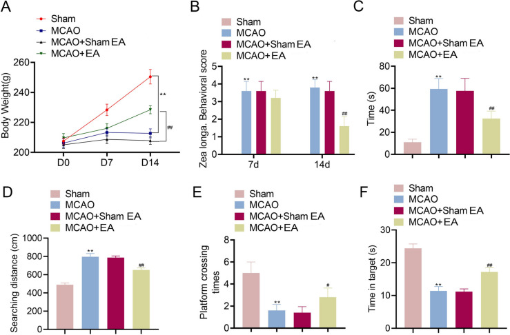
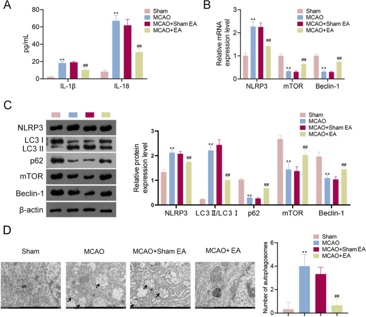
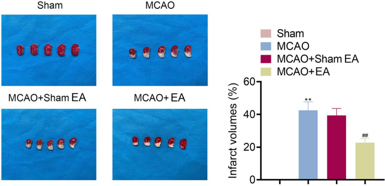
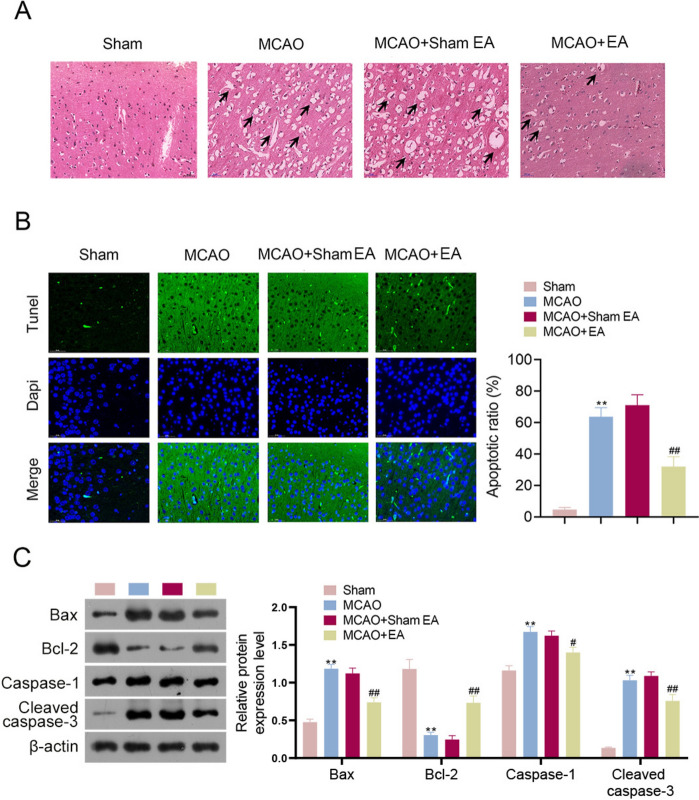
MCAO rats exhibited slower weight gain, reduced mobility, increased infarct size, pathological damage, and apoptosis, confirming successful establishment of the MCAO rat model. Following EA treatment, MCAO rats displayed faster weight gain, improved mobility, and shorter escape latency. EA also reduced the area of cerebral infarction and alleviated pathological damage and apoptosis in MCAO rats. Furthermore, EA downregulated IL-1β, IL-18, NLRP3, and LC3 II/LC3 I expression and upregulated p62, mTOR, and Beclin-1 expression in MCAO rats. EA treatment also decreased the number of autophagosomes in these rats.
EA effectively mitigates post-stroke cognitive impairment by reducing apoptosis, inflammation, and autophagy through the regulation of the mTOR/NLRP3-mediated autophagy-inflammatory pathway, offering valuable therapeutic insights for stroke rehabilitation.
卒中后认知障碍(PSCI)严重降低了卒中患者的生活质量。本研究旨在评估电针对 PSCI 的影响,以及 mTOR/NLRP3 介导的自噬-炎症通路在此过程中的作用。
采用大脑中动脉闭塞(MCAO)法建立大鼠局灶性脑缺血模型。模型成功诱导后,对双侧风池、风府和大椎穴进行电针治疗,于第 15 天采集脑组织样本。采用 Morris 水迷宫试验评估认知功能。采用氯化三苯基四氮唑(TTC)染色定量测定脑梗死体积。通过苏木精-伊红和 TUNEL 染色评估病理变化和细胞凋亡率。检测凋亡、炎症和自噬相关生物标志物,并通过透射电子显微镜观察自噬体。
MCAO 大鼠表现出体重增长缓慢、活动减少、梗死体积增大、病理损伤和细胞凋亡增加,证实 MCAO 大鼠模型建立成功。电针治疗后,MCAO 大鼠体重增长加快,活动能力提高,逃避潜伏期缩短。电针还降低了 MCAO 大鼠的脑梗死面积,减轻了其病理损伤和细胞凋亡。此外,电针还下调了 MCAO 大鼠中 IL-1β、IL-18、NLRP3 和 LC3 II/LC3 I 的表达,上调了 p62、mTOR 和 Beclin-1 的表达。电针治疗还减少了这些大鼠自噬体的数量。
电针通过调节 mTOR/NLRP3 介导的自噬-炎症通路,减少细胞凋亡、炎症和自噬,有效减轻卒中后认知障碍,为卒中康复提供了有价值的治疗思路。