Moraiti Stamatina, Cheong Vee San, Dall'Ara Enrico, Kadirkamanathan Visakan, Bhattacharya Pinaki
Department of Mechanical Engineering, University of Sheffield, Sheffield, United Kingdom.
INSIGNEO Institute for in silico Medicine, University of Sheffield, Sheffield, United Kingdom.
Front Bioeng Biotechnol. 2024 Oct 22;12:1469272. doi: 10.3389/fbioe.2024.1469272. eCollection 2024.
Murine models are used to test the effect of anti-osteoporosis treatments as they replicate some of the bone phenotypes observed in osteoporotic (OP) patients. The effect of disease and treatment is typically described as changes in bone geometry and microstructure over time. Conventional assessment of geometric changes relies on morphometric scalar parameters. However, being correlated with each other, these parameters do not describe separate fractions of variations and offer only a moderate insight into temporal changes.
The current study proposes a novel image-based framework that employs deformable image registration on longitudinal images of bones and Principal Component Analysis (PCA) for improved quantification of geometric effects of OP treatments. This PCA-based model and a novel post-processing of score changes provide orthogonal modes of shape variations temporally induced by a course of treatment (specifically mechanical loading).
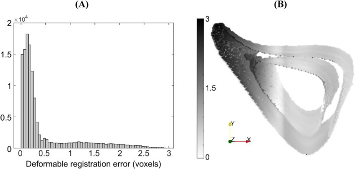
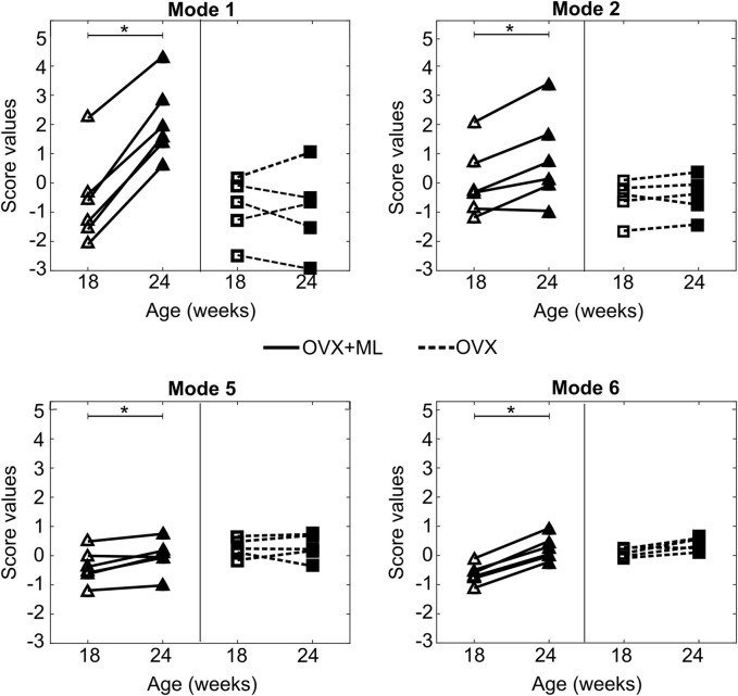
Errors associated with the proposed framework are rigorously quantified and it is shown that the accuracy of deformable image registration in capturing the bone shapes (∼1 voxel = 10.4 μm) is of the same order of magnitude as the relevant state-of-the-art evaluation studies. Applying the framework to longitudinal image data from the midshaft section of ovariectomized mouse tibia, two mutually orthogonal mode shapes are reliably identified to be an effect of treatment. The mode shapes captured changes of the tibia geometry due to the treatment at the anterior crest (maximum of 0.103 mm) and across the tibia midshaft section and the posterior (0.030 mm) and medial (0.024 mm) aspects. These changes agree with those reported previously but are now described in a compact fashion, as a vector field of displacements on the bone surface. The proposed framework enables a more detailed investigation of the effect of disease and treatment on bones in preclinical studies and boosts the precision of such assessments.
小鼠模型用于测试抗骨质疏松治疗的效果,因为它们能复制骨质疏松(OP)患者中观察到的一些骨表型。疾病和治疗的效果通常被描述为随时间变化的骨几何形状和微观结构的改变。传统的几何变化评估依赖于形态计量标量参数。然而,这些参数相互关联,无法描述变化的不同部分,对时间变化的洞察也较为有限。
本研究提出了一种基于图像的新框架,该框架在骨骼纵向图像上采用可变形图像配准,并结合主成分分析(PCA),以改进对OP治疗几何效果的量化。这种基于PCA的模型以及分数变化的新后处理方法提供了由治疗过程(特别是机械负荷)随时间诱导的形状变化的正交模式。
对所提出框架相关的误差进行了严格量化,结果表明,可变形图像配准在捕捉骨形状方面的准确性(约1体素 = 10.4μm)与相关的最新评估研究处于同一数量级。将该框架应用于去卵巢小鼠胫骨中轴段的纵向图像数据,可靠地识别出两个相互正交的模式形状是治疗的效果。这些模式形状捕捉到了由于治疗导致的胫骨前嵴处(最大0.103mm)、整个胫骨中轴段以及后部(0.030mm)和内侧(0.024mm)的几何形状变化。这些变化与先前报道的一致,但现在以一种紧凑的方式描述为骨表面位移的向量场。所提出的框架能够在临床前研究中更详细地研究疾病和治疗对骨骼的影响,并提高此类评估的精度。