Department of Microbiology and Immunology, Dartmouth College Geisel School of Medicine, Lebanon, New Hampshire, USA.
Dartmouth Cancer Center, Dartmouth College Geisel School of Medicine, Lebanon, New Hampshire, USA.
J Immunother Cancer. 2024 Nov 9;12(11):e010170. doi: 10.1136/jitc-2024-010170.
Overcoming immune suppression is a major barrier to eliciting potent CD8 T cell responses against cancer. Treatment with anti-CD4 monoclonal antibody is an effective means for eliminating CD4Foxp3 regulatory (Treg) cells in preclinical models and has also demonstrated efficacy in early clinical trials. However, the underlying basis for treatment efficacy, more specifically the implications of codepleting other CD4-expressing cell compartments in tumor-bearing hosts, is not well understood.
Tumor-bearing mice were treated with anti-CD4 versus other therapies that preserve helper T cell function, and the priming, tissue distribution, and maintenance of tumor antigen-specific CD8 T cells were assessed. Antibody blockade and transgenic mouse models were used to determine the mechanisms of CD8 T cell priming. Single-cell RNA-sequencing (scRNAseq) was used to further characterize CD8 T cells that are primed by anti-CD4 therapy and to identify immunosuppressive CD4 T cell subsets in human melanoma following immune checkpoint blockade (ICB).
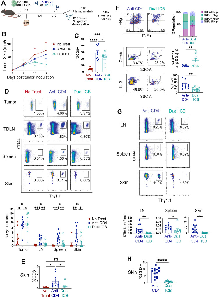
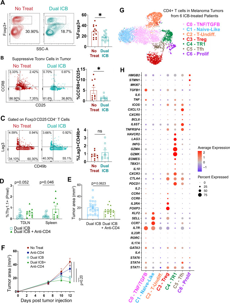
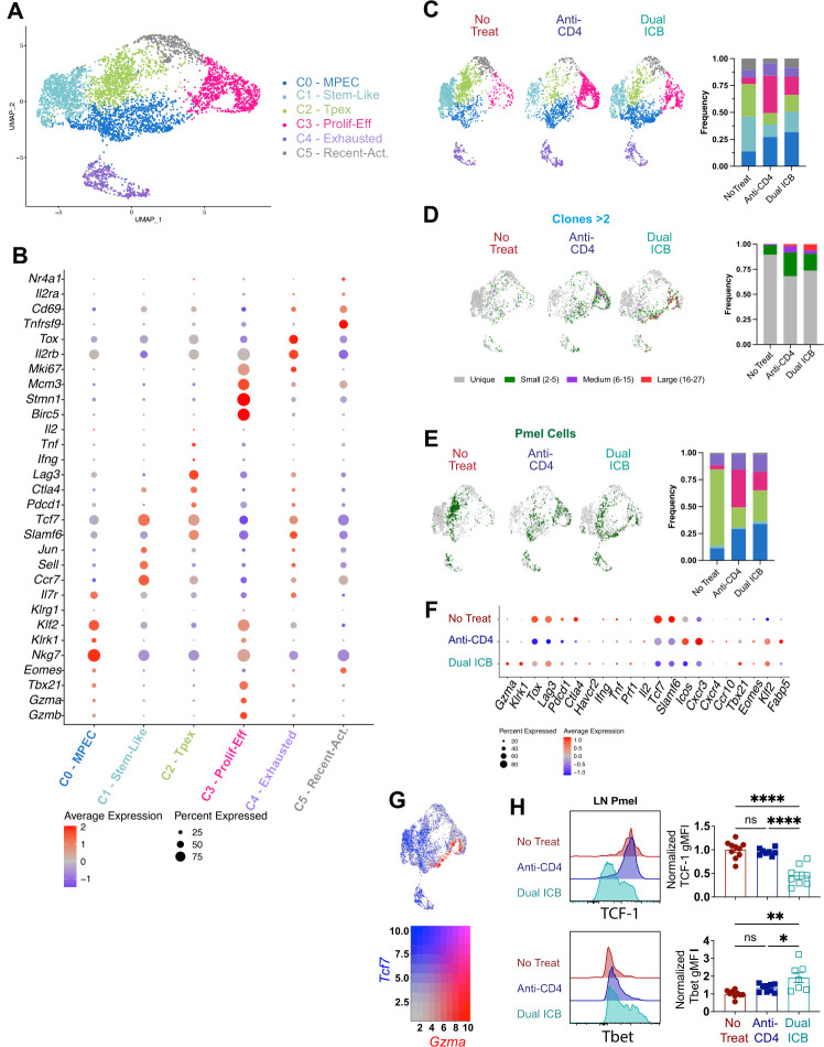
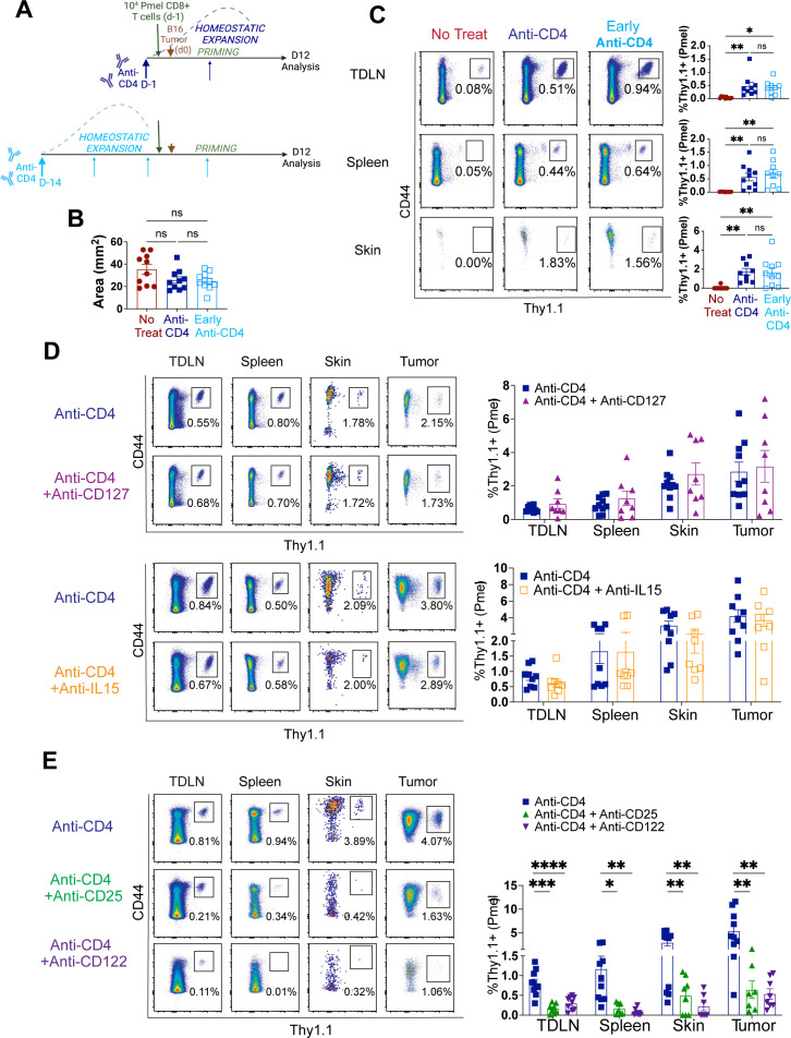
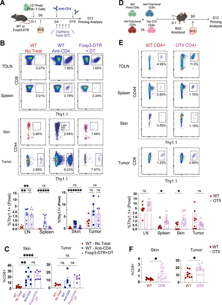
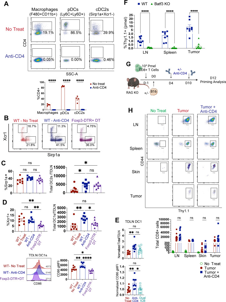
Comparing anti-CD4 to dual ICB therapy, we show that anti-CD4 facilitates more robust priming of TCF-1, IL-2-producing, tumor-specific CD8 T cells that disseminate to tissues and form memory. By decoupling priming from homeostatic proliferation and associated cytokines, we find that anti-CD4 functions independently of creating homeostatic space for CD8 T cells. We also show that depletion of CD4-expressing antigen-presenting cell subsets is not required for anti-CD4 efficacy. Instead, robust tumor-specific CD8 T cell priming and memory generation required the removal of total antigen-specific CD4 T cells, including both Tregs and CD4 Foxp3-negative conventional (Tconv) cells. In particular, the elimination of CD4 Tconv cells was necessary for the accumulation and maturation of conventional type-1 dendritic cells in tumor-draining LNs, which were required for CD8 T cell priming. Accordingly, anti-CD4 treatment restored CD8 T cell responses in mice cotreated with dual ICB. scRNAseq of melanoma tumors from patients who received ICB revealed the presence of Tr1 and Treg subsets, as well as CD4 Tconv subsets that lacked clear transcriptional evidence of helper differentiation.
These findings underscore the underappreciated benefit of depleting CD4 Tconv cells to promote systemic primary and memory CD8 T cell responses against cancer.
克服免疫抑制是引发针对癌症的有效 CD8 T 细胞反应的主要障碍。抗 CD4 单克隆抗体的治疗是消除临床前模型中 CD4Foxp3 调节性(Treg)细胞的有效手段,并且在早期临床试验中也显示出疗效。然而,治疗效果的基础,更具体地说,在荷瘤宿主中耗竭其他表达 CD4 的细胞区室的影响,尚不清楚。
用抗 CD4 治疗荷瘤小鼠,与保留辅助 T 细胞功能的其他疗法进行比较,评估肿瘤抗原特异性 CD8 T 细胞的启动、组织分布和维持。抗体阻断和转基因小鼠模型用于确定 CD8 T 细胞启动的机制。单细胞 RNA 测序(scRNAseq)用于进一步描述抗 CD4 治疗诱导的 CD8 T 细胞,并鉴定人黑色素瘤在免疫检查点阻断(ICB)后免疫抑制性 CD4 T 细胞亚群。
与双重 ICB 治疗相比,我们发现抗 CD4 更有利于 TCF-1、IL-2 产生、肿瘤特异性 CD8 T 细胞的有效启动,这些细胞会扩散到组织中并形成记忆。通过将启动与稳态增殖和相关细胞因子分离,我们发现抗 CD4 独立于为 CD8 T 细胞创造稳态空间而发挥作用。我们还表明,耗竭表达 CD4 的抗原呈递细胞亚群不是抗 CD4 疗效所必需的。相反,强烈的肿瘤特异性 CD8 T 细胞启动和记忆生成需要去除所有抗原特异性 CD4 T 细胞,包括 Tregs 和 CD4 Foxp3 阴性常规(Tconv)细胞。特别是,CD4 Tconv 细胞的消除对于肿瘤引流淋巴结中常规 1 型树突状细胞的积累和成熟是必要的,这是 CD8 T 细胞启动所必需的。因此,抗 CD4 治疗恢复了接受双重 ICB 联合治疗的小鼠的 CD8 T 细胞反应。接受 ICB 的黑色素瘤肿瘤的 scRNAseq 揭示了 Tr1 和 Treg 亚群的存在,以及缺乏明显辅助分化转录证据的 CD4 Tconv 亚群。
这些发现强调了耗竭 CD4 Tconv 细胞以促进针对癌症的全身原发性和记忆性 CD8 T 细胞反应的潜在益处。