Innate Immunity Laboratory, Faculty of Advanced Life Science, Hokkaido University, Sapporo, Hokkaido, Japan.
Innate Immunity Laboratory, Graduate School of Life Science, Hokkaido University, Sapporo, Hokkaido, Japan.
PLoS One. 2024 Nov 15;19(11):e0313213. doi: 10.1371/journal.pone.0313213. eCollection 2024.
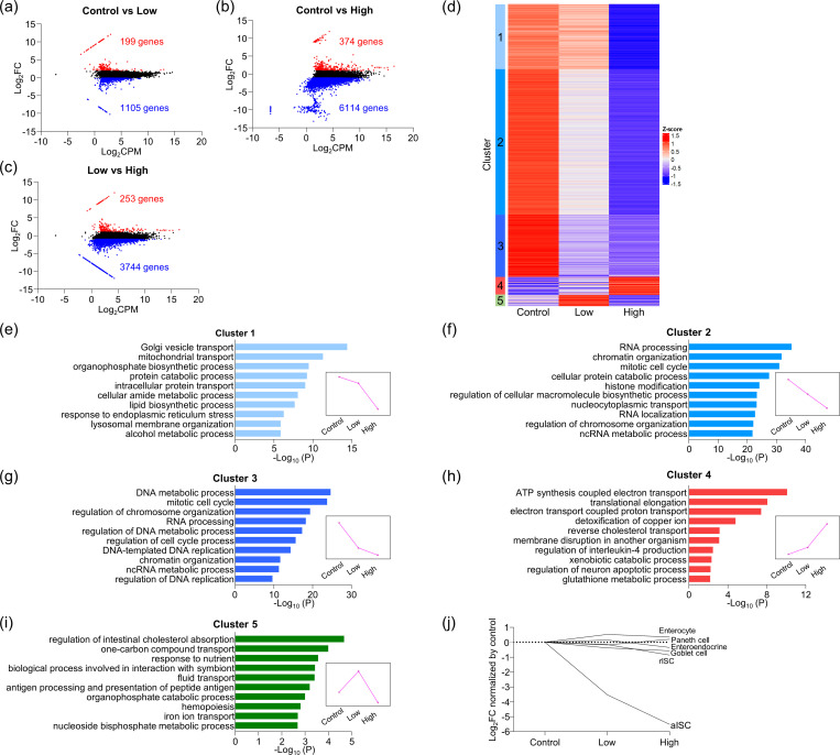
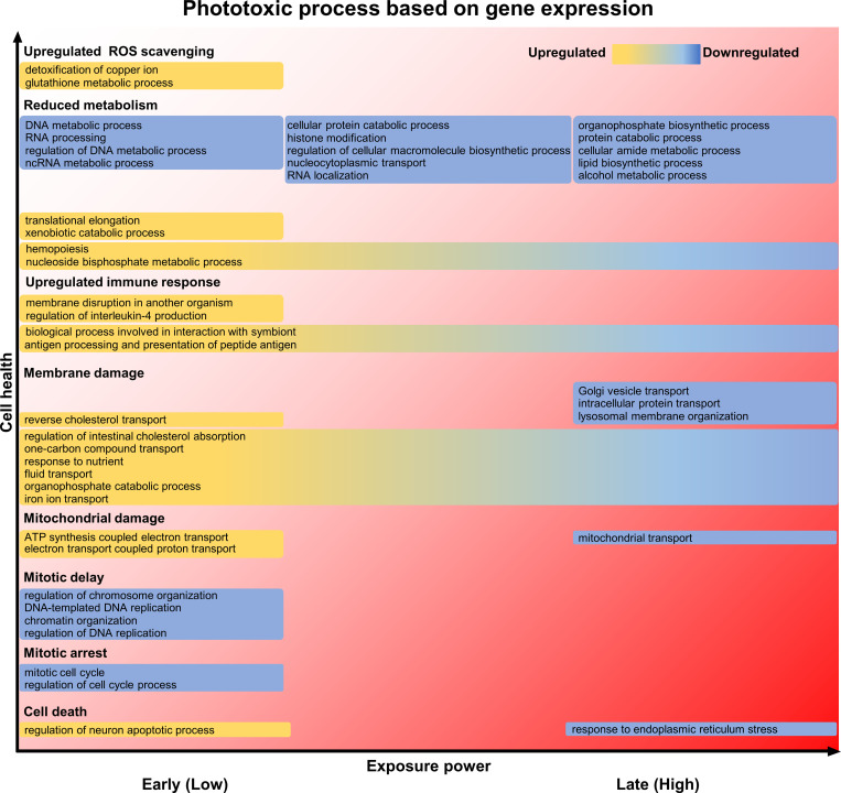
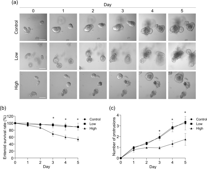
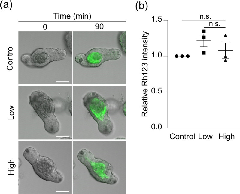
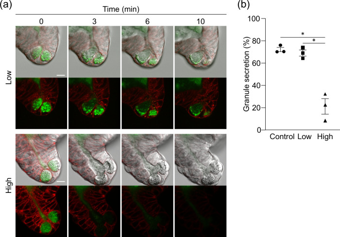
Live imaging visualizes the structure, dynamics, and function of cells and tissues to reveal the molecular mechanisms, and has contributed to the advancement of life science. In live imaging, it has been well known that there is a trade-off between higher-resolution analysis and cell damage caused by light illumination, i.e., phototoxicity. However, despite the risk of unknowingly distorting experimental results, phototoxicity is an unresolved issue in live imaging because overall consequences occurring inside cells due to phototoxicity remains unknown. Here, we determined the molecular process of phototoxicity-induced cell damage systematically under low- and high-dose light illumination conditions by analyzing differential gene expression using RNA-sequencing in a three-dimensional organoid of small intestinal epithelial cells, enteroid. The low-dose light illumination already induced various abnormalities in functional molecules involved in the response to reactive oxygen species generated by the excitation of fluorescent dyes, intracellular metabolism, mitosis, immune responses, etc., at mRNA expression level. Together with the behavior toward apoptosis caused by high-dose light illumination, the light dose-dependent progression of intracellular damage was revealed. About visible impairment of intestinal epithelial function, failures in both the structure-forming ability of enteroids and Paneth cell granule secretion were observed under high-dose light illumination, while the drug efflux was not disturbed despite abnormal drug efflux transporter mRNA expression. Based on the gene expression profiles, we comprehensively clarified phenomena in the cells at mRNA level that cannot be recognized both morphologically and functionally during live imaging, further providing a new insight into the risk of phototoxicity. This study warns from the aspect of mRNA expression that awareness of phototoxic artifacts is needed when analyzing cellular function and the mechanism in live imaging.
实时成像可可视化细胞和组织的结构、动态和功能,揭示分子机制,为生命科学的发展做出了贡献。在实时成像中,人们已经清楚地认识到,更高的分辨率分析与光照射引起的细胞损伤(即光毒性)之间存在权衡。然而,尽管存在不自觉地扭曲实验结果的风险,但光毒性仍然是实时成像中的一个未解决的问题,因为由于光毒性而导致细胞内发生的总体后果仍然未知。在这里,我们通过在三维小肠上皮细胞类器官(肠类器官)中使用 RNA 测序分析差异基因表达,系统地确定了在低剂量和高剂量光照射条件下光毒性诱导的细胞损伤的分子过程。低剂量光照射已经在涉及对荧光染料激发产生的活性氧的反应、细胞内代谢、有丝分裂、免疫反应等的功能分子的 mRNA 表达水平上诱导了各种异常。与高剂量光照射引起的细胞凋亡行为一起,揭示了细胞内损伤的光剂量依赖性进展。关于肠上皮功能的明显损伤,在高剂量光照射下,肠类器官的结构形成能力和潘氏细胞颗粒分泌都出现了失败,而尽管药物外排转运体的 mRNA 表达异常,药物外排仍未受到干扰。基于基因表达谱,我们全面阐明了在实时成像中,细胞在形态学和功能上都无法识别的 mRNA 水平上的现象,进一步深入了解了光毒性的风险。这项研究从 mRNA 表达的角度警告说,在实时成像中分析细胞功能和机制时,需要意识到光毒性的人工制品。