National Institute of Neurological Disorders and Stroke, National Institutes of Health, Bethesda, MD, United States; Department of Neurology, University of California Los Angeles David Geffen School of Medicine, Los Angeles, CA, United States.
National Institute of Neurological Disorders and Stroke, National Institutes of Health, Bethesda, MD, United States.
Neuroimage Clin. 2024;44:103706. doi: 10.1016/j.nicl.2024.103706. Epub 2024 Nov 13.
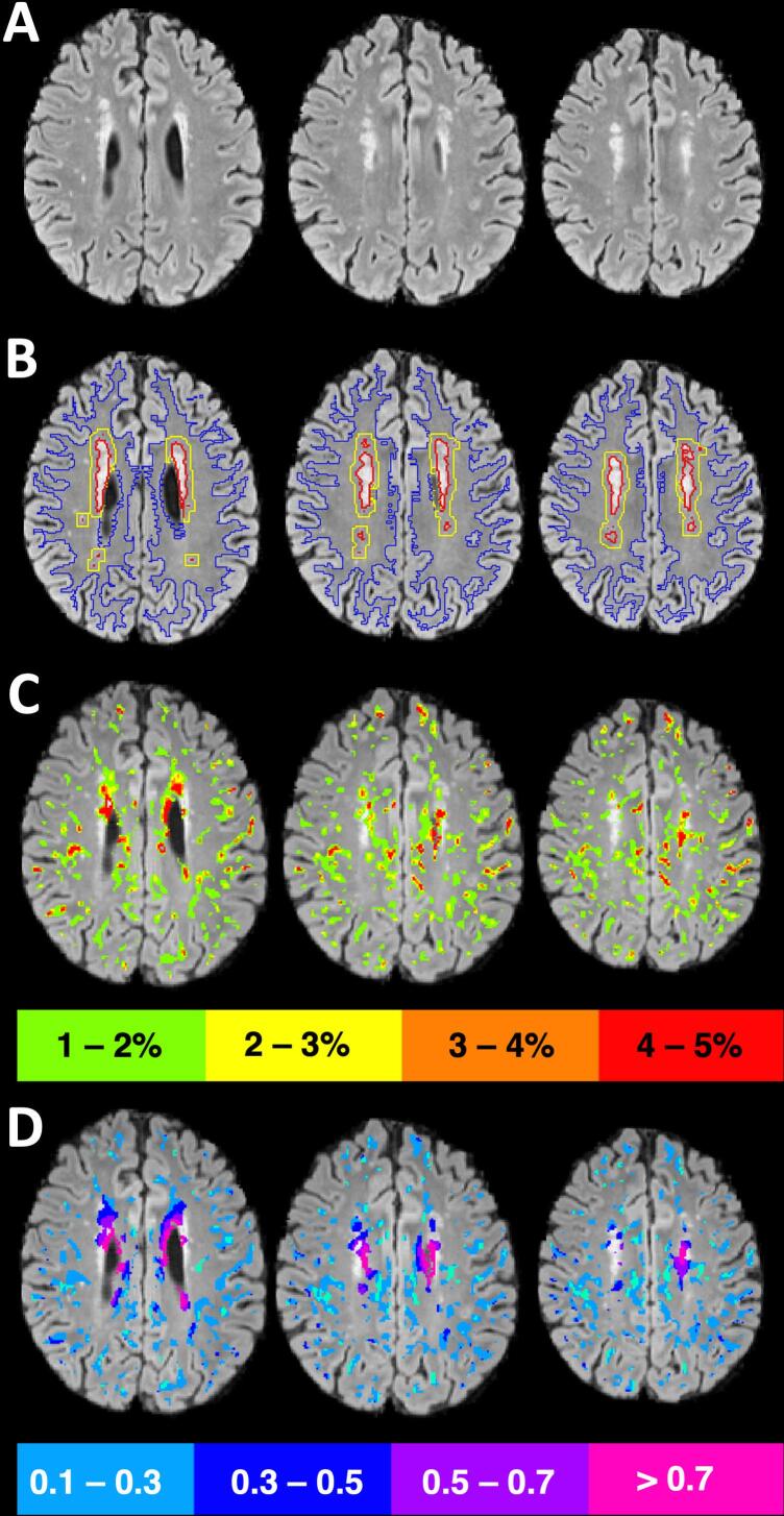
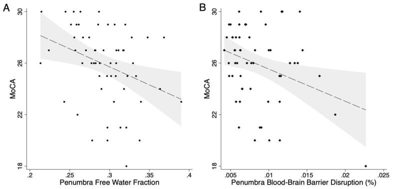
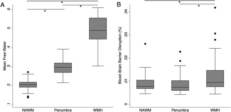
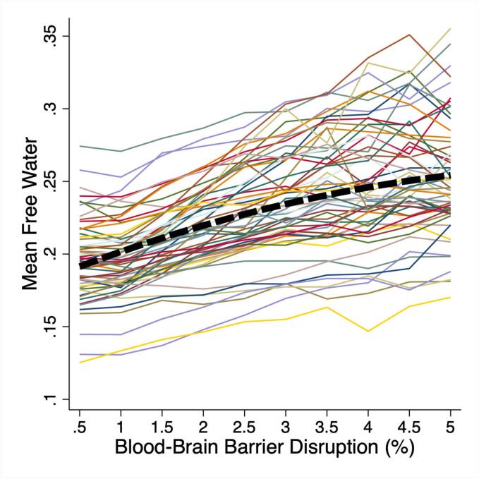
Progression of cerebral small vessel disease (CSVD) is associated with cognitive decline. Blood-brain barrier disruption (BBBD) and fluid extravasation to the interstitial space may contribute to progression of white matter hyperintensities (WMH). We hypothesized that increased free water (FW) would colocalize with BBBD and relate to cognitive performance. Patients with ischemic stroke/TIA at least 3 months prior with at least early confluent WMH were studied cross-sectionally with the Montreal Cognitive Assessment (MoCA), diffusion tensor imaging, and dynamic susceptibility contrast imaging. White matter (WM) was segmented into WMH, WMH penumbra, and normal appearing white matter (NAWM). Colocalization of elevated FW and BBBD and their associations with MoCA performance were evaluated. 58 patients were included (mean age 69, 36 % female). Higher BBBD colocalized with elevated FW. Elevated FW in all white matter, NAWM, WMH penumbra, and WMH lesions was associated with lower MoCA score. Increased BBBD in all WM, NAWM, and WMH penumbra was associated with lower MoCA. In WMH penumbra, both elevated FW and increased BBBD were independently associated with lower MoCA. We found agreement between 2 different biomarkers implicated in the pathogenesis of CSVD that independently demonstrated association with cognitive performance when measured in the area of postulated disease activity.
脑小血管病(CSVD)的进展与认知能力下降有关。血脑屏障破坏(BBBD)和液体外渗到细胞间隙可能导致脑白质高信号(WMH)的进展。我们假设增加的自由水(FW)会与 BBBD 共定位,并与认知表现相关。对至少 3 个月前发生缺血性卒中和 TIA 且至少存在早期融合性 WMH 的患者进行了横断面研究,研究方法包括蒙特利尔认知评估(MoCA)、弥散张量成像和动态对比增强磁共振成像。将脑白质(WM)分为 WMH、WMH 半影区和正常表现的脑白质(NAWM)。评估了升高的 FW 与 BBBD 的共定位及其与 MoCA 表现的关系。共纳入 58 例患者(平均年龄 69 岁,36%为女性)。升高的 BBBD 与升高的 FW 共定位。所有脑白质、NAWM、WMH 半影区和 WMH 病变中的升高 FW 与 MoCA 评分降低相关。所有 WM、NAWM 和 WMH 半影区中的升高 BBBD 与 MoCA 降低相关。在 WMH 半影区中,升高的 FW 和增加的 BBBD 均与 MoCA 降低独立相关。我们发现,两种不同的生物标志物均与 CSVD 的发病机制有关,当在假定的疾病活动区域测量时,这两种生物标志物均与认知表现独立相关,这两种标志物之间存在一致性。