• 文献检索
  • 文档翻译
  • 深度研究
  • 学术资讯
  • Suppr Zotero 插件Zotero 插件
  • 邀请有礼
  • 套餐&价格
  • 历史记录
应用&插件
Suppr Zotero 插件Zotero 插件浏览器插件Mac 客户端Windows 客户端微信小程序
定价
高级版会员购买积分包购买API积分包
服务
文献检索文档翻译深度研究API 文档MCP 服务
关于我们
关于 Suppr公司介绍联系我们用户协议隐私条款
关注我们

Suppr 超能文献

核心技术专利:CN118964589B侵权必究
粤ICP备2023148730 号-1Suppr @ 2026

文献检索

告别复杂PubMed语法,用中文像聊天一样搜索,搜遍4000万医学文献。AI智能推荐,让科研检索更轻松。

立即免费搜索

文件翻译

保留排版,准确专业,支持PDF/Word/PPT等文件格式,支持 12+语言互译。

免费翻译文档

深度研究

AI帮你快速写综述,25分钟生成高质量综述,智能提取关键信息,辅助科研写作。

立即免费体验

一例可切除的单结节肝内胆管腺瘤病例。

A Case of Resectable Single-Nodule Intrahepatic Bile Duct Adenoma.

作者信息

Okano Hiroshi, Asakawa Hiroki, Mukai Katsumi, Nishimura Akira, Hamada Takashi, Asakawa Kana, Baba Youichirou, Murata Tetsuya

机构信息

Gastroenterology, Suzuka General Hospital, Suzuka, JPN.

Gastroenterology, Suzuka general hospital, Suzuka, JPN.

出版信息

Cureus. 2024 Oct 16;16(10):e71656. doi: 10.7759/cureus.71656. eCollection 2024 Oct.

DOI:10.7759/cureus.71656
PMID:39553064
原文链接:https://pmc.ncbi.nlm.nih.gov/articles/PMC11567730/
Abstract

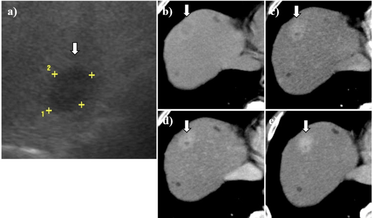
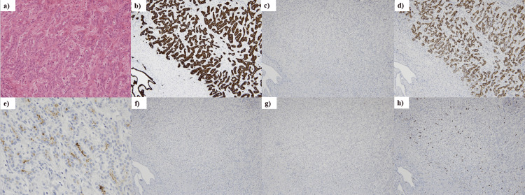
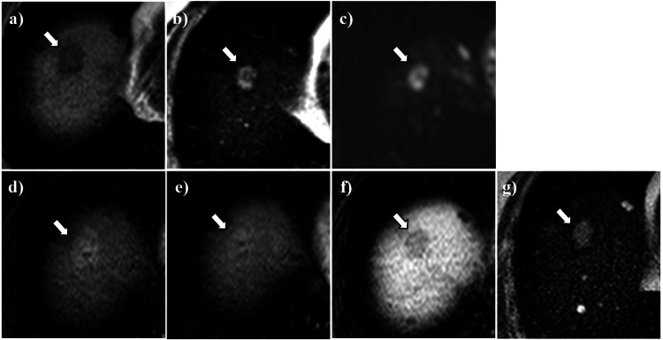
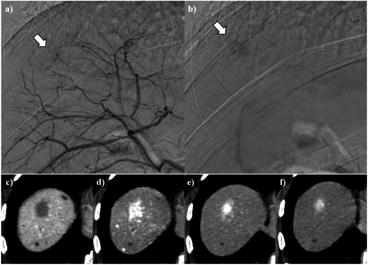
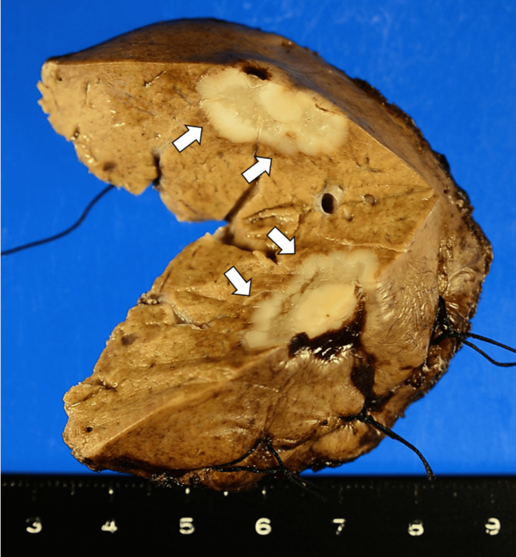
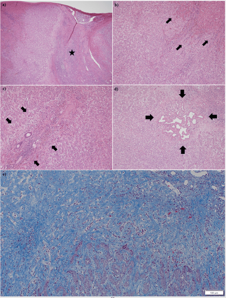
A 70-year-old man was incidentally diagnosed with a single hepatic mass lesion in his right hepatic lobe during a computed tomography scan. The lesion exhibited consistent enhancement with contrast agents on computed tomography (CT), magnetic resonance imaging (MRI), and hepatic arterial angiography. While a definitive diagnosis could not be made preoperatively, the lesion was surgically resected due to its slight enlargement over two months, suggesting a potential malignancy. Pathological examination revealed the lesion to be a bile duct adenoma (BDA). The BDA was characterized by dense proliferative small gland cavities containing several to dozens of cells. Immunohistochemical staining showed positive CK7 and negative p53. The patient remains alive and free of recurrence five years after hepatectomy. Although BDAs are rare benign hepatic tumors, they carry a risk of harboring or developing malignant tissue, such as cholangiocarcinoma. Therefore, BDAs or lesions suspicious of BDA should be surgically resected or closely monitored.

摘要

一名70岁男性在计算机断层扫描时偶然发现右肝叶有一个单发肝脏肿块病变。该病变在计算机断层扫描(CT)、磁共振成像(MRI)和肝动脉血管造影中表现出与造影剂一致的强化。虽然术前未能做出明确诊断,但由于该病变在两个月内略有增大,提示可能为恶性,因此进行了手术切除。病理检查显示该病变为胆管腺瘤(BDA)。BDA的特征是密集增生的小腺腔,含有几个到几十个细胞。免疫组织化学染色显示CK7阳性,p53阴性。肝切除术后五年,患者仍然存活且无复发。虽然BDA是罕见的肝脏良性肿瘤,但它们有 harboring 或发展为恶性组织(如胆管癌)的风险。因此,BDA或疑似BDA的病变应进行手术切除或密切监测。 (注:原英文中“harboring”这个词在这里结合语境推测可能是“隐匿”之类意思,但直接翻译不好处理,这里保留英文供你参考确认准确意思。)

https://cdn.ncbi.nlm.nih.gov/pmc/blobs/8e10/11567730/49fcc5b8950d/cureus-0016-00000071656-i06.jpg
https://cdn.ncbi.nlm.nih.gov/pmc/blobs/8e10/11567730/d3f6113b0340/cureus-0016-00000071656-i01.jpg
https://cdn.ncbi.nlm.nih.gov/pmc/blobs/8e10/11567730/699eedd9a5d9/cureus-0016-00000071656-i02.jpg
https://cdn.ncbi.nlm.nih.gov/pmc/blobs/8e10/11567730/03d71878586e/cureus-0016-00000071656-i03.jpg
https://cdn.ncbi.nlm.nih.gov/pmc/blobs/8e10/11567730/be13cb1555e5/cureus-0016-00000071656-i04.jpg
https://cdn.ncbi.nlm.nih.gov/pmc/blobs/8e10/11567730/01138418b2cc/cureus-0016-00000071656-i05.jpg
https://cdn.ncbi.nlm.nih.gov/pmc/blobs/8e10/11567730/49fcc5b8950d/cureus-0016-00000071656-i06.jpg

相似文献

1
A Case of Resectable Single-Nodule Intrahepatic Bile Duct Adenoma.一例可切除的单结节肝内胆管腺瘤病例。
Cureus. 2024 Oct 16;16(10):e71656. doi: 10.7759/cureus.71656. eCollection 2024 Oct.
2
Intrahepatic bile duct adenoma (peribiliary gland hamartoma): a case report and review of literature.肝内胆管腺瘤(胆管周围腺错构瘤):一例病例报告及文献复习
Int J Clin Exp Pathol. 2015 May 1;8(5):5908-13. eCollection 2015.
3
Intrahepatic bile duct adenoma in a patient with gastric cancer.一名胃癌患者的肝内胆管腺瘤
Int Cancer Conf J. 2018 Sep 12;8(1):7-11. doi: 10.1007/s13691-018-0345-y. eCollection 2019 Jan.
4
Intrahepatic Cholangiocarcinoma Coexisting With Multiple Bile Duct Adenoma Treated as Liver Metastasis from a Pancreatic Neuroendocrine Tumor.肝内胆管细胞癌合并多发胆管腺瘤被误诊为胰腺神经内分泌肿瘤肝转移。
Anticancer Res. 2021 Oct;41(10):5249-5254. doi: 10.21873/anticanres.15344.
5
Magnetic Resonance Imaging Findings of an Intrahepatic Bile Duct Adenoma: A Case Report.肝内胆管腺瘤的磁共振成像表现:一例报告
Cureus. 2022 Jul 20;14(7):e27082. doi: 10.7759/cureus.27082. eCollection 2022 Jul.
6
Imaging features of bile duct adenoma: case series and review of literature.胆管腺瘤的影像学特征:病例系列及文献复习。
Diagn Interv Radiol. 2018 Sep;24(5):249-254. doi: 10.5152/dir.2018.18036.
7
Adenomyoma of the common hepatic duct mimicking bile duct cancer: report of a case.酷似胆管癌的肝总管腺肌瘤:病例报告
Surg Today. 2007;37(7):608-11. doi: 10.1007/s00595-006-3468-2. Epub 2007 Jun 26.
8
Magnetic Resonance Imaging Findings of Intrahepatic Bile Duct Adenoma: A Report of 4 Cases.肝内胆管腺瘤的磁共振成像表现:4例报告
J Comput Assist Tomogr. 2015 Sep-Oct;39(5):747-51. doi: 10.1097/RCT.0000000000000286.
9
Bile duct adenoma: imaging features and radiologic-pathologic correlation.胆管腺瘤:影像学特征及放射学-病理学相关性
Jpn J Radiol. 2020 Jun;38(6):561-571. doi: 10.1007/s11604-020-00938-0. Epub 2020 Mar 3.
10
Bile duct adenoma: a case report and literature review.胆管腺瘤:一例病例报告及文献综述
World J Surg Oncol. 2014 Apr 26;12:125. doi: 10.1186/1477-7819-12-125.

本文引用的文献

1
ACG Clinical Guideline: Focal Liver Lesions.ACG 临床指南:局灶性肝脏病变。
Am J Gastroenterol. 2024 Jul 1;119(7):1235-1271. doi: 10.14309/ajg.0000000000002857. Epub 2024 Jan 26.
2
High expression of autotaxin is associated with poor recurrence-free survival in cholangiocarcinoma.自分泌运动因子的高表达与胆管癌患者无复发生存期短相关。
Hepatol Res. 2024 Sep;54(9):817-826. doi: 10.1111/hepr.14031. Epub 2024 Mar 2.
3
Bile duct adenoma and small-sized small duct type intrahepatic cholangiocarcinoma show distinct differences in genetic alterations, expression of IMP3 and EZH2 and stromal and inflammatory components.
胆管腺瘤和小胆管型肝内胆管癌在遗传改变、IMP3 和 EZH2 的表达、基质和炎症成分方面存在明显差异。
Histopathology. 2023 Aug;83(2):298-309. doi: 10.1111/his.14932. Epub 2023 May 4.
4
Focal Benign Liver Lesions and Their Diagnostic Pitfalls.肝脏局灶性良性病变及其诊断陷阱。
Radiol Clin North Am. 2022 Sep;60(5):755-773. doi: 10.1016/j.rcl.2022.05.005. Epub 2022 Jul 4.
5
Incidental Radiology Findings on Computed Tomography Studies in Emergency Department Patients: A Systematic Review and Meta-Analysis.急诊科患者计算机断层扫描研究中的偶然放射学发现:系统评价和荟萃分析。
Ann Emerg Med. 2022 Sep;80(3):243-256. doi: 10.1016/j.annemergmed.2022.03.027. Epub 2022 Jun 16.
6
Bile duct adenoma: rare benign liver tumour.胆管腺瘤:罕见的肝脏良性肿瘤。
Singapore Med J. 2022 Dec;63(12):769-770. doi: 10.11622/smedj.2021101. Epub 2021 Sep 21.
7
Bile duct adenoma may be a precursor lesion of small duct type intrahepatic cholangiocarcinoma.胆管腺瘤可能是小胆管型肝内胆管癌的前驱病变。
Histopathology. 2021 Jan;78(2):310-320. doi: 10.1111/his.14222. Epub 2020 Sep 15.
8
Serum ATX as a novel biomarker for breast cancer.血清自分泌运动因子作为乳腺癌的一种新型生物标志物。
Medicine (Baltimore). 2019 Mar;98(13):e14973. doi: 10.1097/MD.0000000000014973.
9
Cholangiocarcinoma: Classification, Histopathology and Molecular Carcinogenesis.胆管癌:分类、组织病理学和分子发生机制。
Pathol Oncol Res. 2020 Jan;26(1):3-15. doi: 10.1007/s12253-018-0491-8. Epub 2018 Nov 17.
10
Imaging features of bile duct adenoma: case series and review of literature.胆管腺瘤的影像学特征:病例系列及文献复习。
Diagn Interv Radiol. 2018 Sep;24(5):249-254. doi: 10.5152/dir.2018.18036.