Tran Phillip, Schneider Nils, Cho Jaden, Parrish Todd B, Tate Matthew C, Iorga Michael
Departments of Radiology, Northwestern University, Chicago, Illinois.
Departments of Biomedical Engineering, Northwestern University, Chicago, Illinois.
J Neurosurg Case Lessons. 2024 Nov 18;8(21). doi: 10.3171/CASE24549.
The leading method of identifying critical functional regions during brain tumor resection is direct electrical stimulation (DES). In awake craniotomy patients, DES employs electric current to induce functional responses or task inhibition. In contrast, thermography uses infrared imaging to detect regions of increased blood flow from patient tasks, inferring the location of functional activity similarly to blood oxygen level-dependent (BOLD) functional magnetic resonance imaging (fMRI). DES seldom produces no detectable response, but the case herein is an example featuring the subsequent use of thermography.
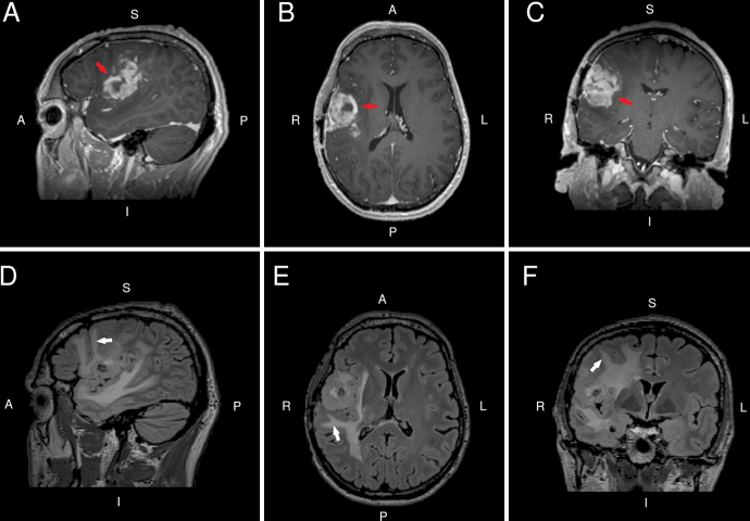
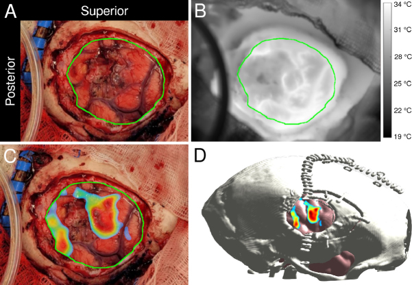
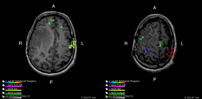
The authors present the case of a 40-year-old male in whom awake craniotomy DES for high-grade glioma re-resection produced no detectable response at the upper levels of tolerated current amplitude. Following inconclusive DES, infrared thermography was performed with a lip-pursing task, and face motor activation was thermally detected in regions corroborated by both preoperative BOLD fMRI and literature on BOLD fMRI face motor mapping.
The lack of a detectable DES response was attributed to significant peritumoral edema, as evidenced by preoperative fluid-attenuated inversion recovery MRI. Findings indicate that infrared thermography overcomes the limitations of DES in an extensive edema setting and that thermography offers a useful complement to standard cortical mapping protocols for resection planning. https://thejns.org/doi/10.3171/CASE24549.
在脑肿瘤切除术中识别关键功能区的主要方法是直接电刺激(DES)。在清醒开颅手术患者中,DES利用电流诱发功能反应或任务抑制。相比之下,热成像使用红外成像来检测患者任务引起的血流增加区域,类似于血氧水平依赖(BOLD)功能磁共振成像(fMRI)推断功能活动的位置。DES很少不产生可检测到的反应,但本文所述病例是随后使用热成像的一个例子。
作者介绍了一名40岁男性的病例,该患者因高级别胶质瘤再次切除进行清醒开颅DES时,在耐受电流幅度的上限未产生可检测到的反应。在DES结果不明确后,进行了唇部收缩任务的红外热成像,在术前BOLD fMRI和关于BOLD fMRI面部运动映射的文献证实的区域热检测到面部运动激活。
术前液体衰减反转恢复MRI显示,未检测到DES反应归因于显著的瘤周水肿。研究结果表明,红外热成像克服了DES在广泛水肿情况下的局限性,并且热成像为切除计划的标准皮质映射方案提供了有用的补充。https://thejns.org/doi/10.3171/CASE24549。