Pleouras Dimitrios S, Loukas Vasileios S, Karanasiou Georgia, Katsouras Christos, Semertzioglou Arsen, Moulas Anargyros N, Michalis Lambros K, Fotiadis Dimitrios I
Unit of Medical Technology and Intelligent Information Systems, Department of Materials Science and EngineeringUniversity of Ioannina GR45110 Ioannina Greece.
Biomedical Research Institute - FORTHUniversity Campus of Ioannina GR45110 Ioannina Greece.
IEEE Open J Eng Med Biol. 2024 May 16;6:1-9. doi: 10.1109/OJEMB.2024.3402057. eCollection 2025.
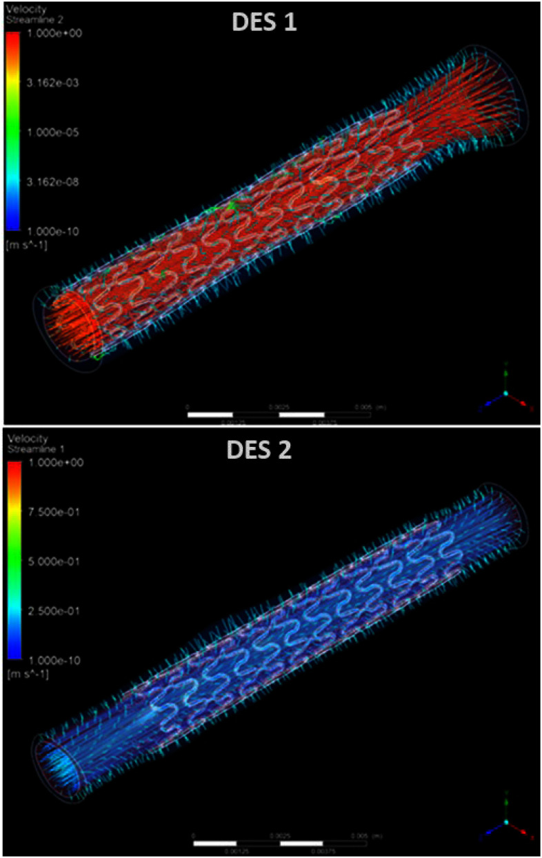
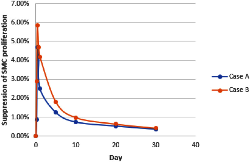
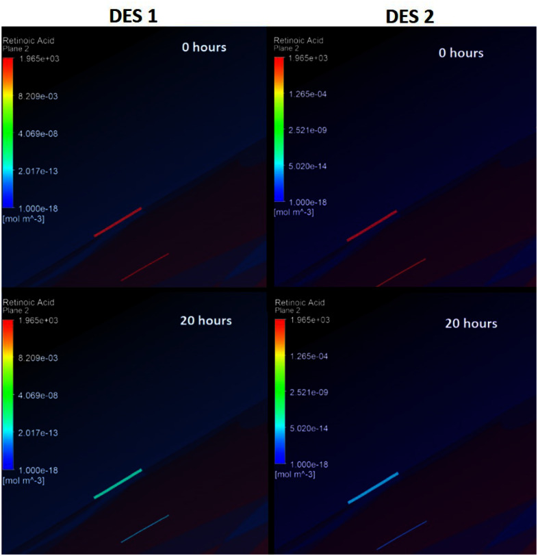
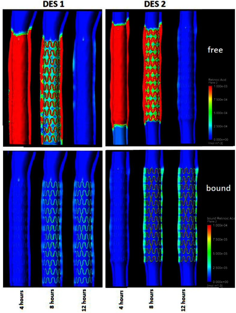
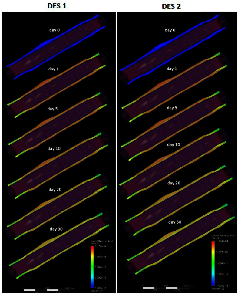
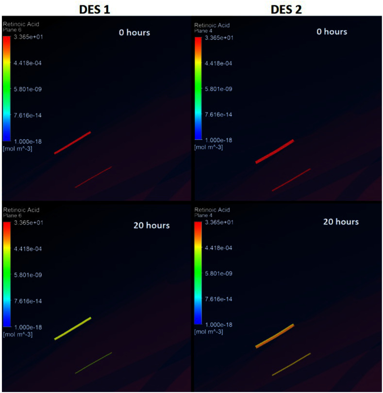
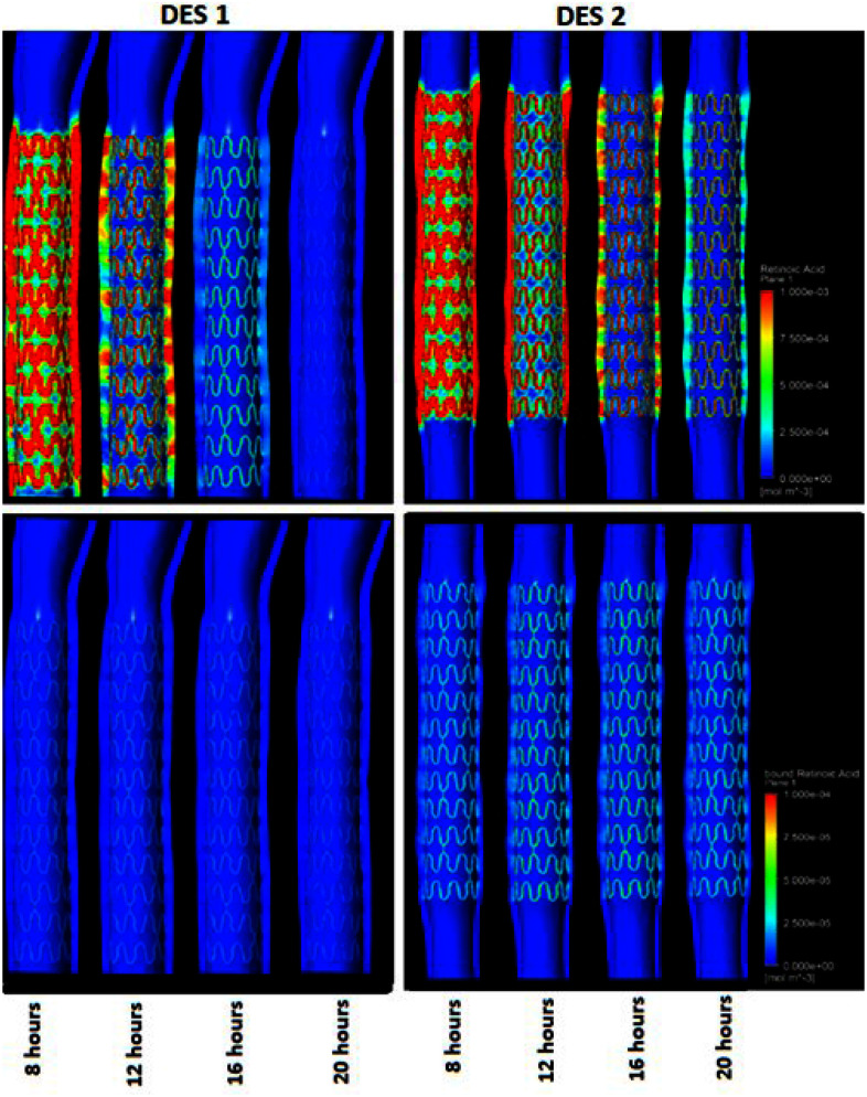
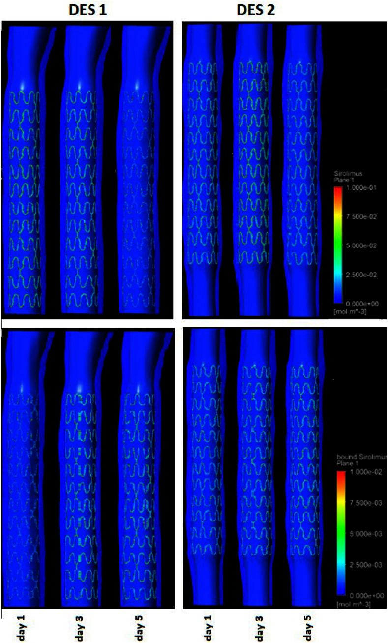
In this work, a methodology for the in-silico evaluation of drug eluting stents (DES) is presented. A stent model developed by Rontis S.A. has been employed. For modeling purposes two different stent parts have been considered: the metal core and the coating. For the arterial models, we used animal specific imaging data and realistic geometries were reconstructed which were used as input to the drug-delivery model. More specifically, optical coherence tomography (OCT) imaging data from two coney iliac arterial segments were 3D reconstructed, and the preprocessed 3D stent was deployed in-silico. The deformed geometries of the in-silico deployed stents and the dilated arterial segments were used as input to the drug elution model. The same reconstructed arteries were used in three different cases: (i) Case A. The coatings contain retinoic acid at an initial concentration 49.2% w/w. (ii) Case B. The coatings contain retinoic acid at an initial concentration 1% w/w. (iii) Case C. The coatings contain sirolimus at an initial concentration 0.85% w/w. In each case, two different coatings were examined: (a) polylactic acid and (b) polylactic-co-glycolic acid. The results proved that retinoic acid is a very promising drug candidate for DES due to its binding time to the smooth muscle cells of the arterial wall that exceeds the corresponding time of sirolimus, while being non-toxic to the smooth muscle cells.
在这项工作中,提出了一种用于药物洗脱支架(DES)计算机模拟评估的方法。采用了由Rontis S.A.开发的支架模型。出于建模目的,考虑了两种不同的支架部件:金属芯和涂层。对于动脉模型,我们使用了动物特异性成像数据,并重建了逼真的几何形状,将其用作药物输送模型的输入。更具体地说,对来自两个兔髂动脉段的光学相干断层扫描(OCT)成像数据进行了三维重建,并在计算机模拟中部署了预处理后的三维支架。计算机模拟部署的支架和扩张动脉段的变形几何形状被用作药物洗脱模型的输入。在三种不同情况下使用相同重建的动脉:(i)情况A。涂层含有初始浓度为49.2%w/w的视黄酸。(ii)情况B。涂层含有初始浓度为1%w/w的视黄酸。(iii)情况C。涂层含有初始浓度为0.85%w/w的西罗莫司。在每种情况下,检查了两种不同的涂层:(a)聚乳酸和(b)聚乳酸-乙醇酸共聚物。结果证明,视黄酸是一种非常有前景的DES药物候选物,因为它与动脉壁平滑肌细胞的结合时间超过了西罗莫司的相应时间,同时对平滑肌细胞无毒。