• 文献检索
  • 文档翻译
  • 深度研究
  • 学术资讯
  • Suppr Zotero 插件Zotero 插件
  • 邀请有礼
  • 套餐&价格
  • 历史记录
应用&插件
Suppr Zotero 插件Zotero 插件浏览器插件Mac 客户端Windows 客户端微信小程序
定价
高级版会员购买积分包购买API积分包
服务
文献检索文档翻译深度研究API 文档MCP 服务
关于我们
关于 Suppr公司介绍联系我们用户协议隐私条款
关注我们

Suppr 超能文献

核心技术专利:CN118964589B侵权必究
粤ICP备2023148730 号-1Suppr @ 2026

文献检索

告别复杂PubMed语法,用中文像聊天一样搜索,搜遍4000万医学文献。AI智能推荐,让科研检索更轻松。

立即免费搜索

文件翻译

保留排版,准确专业,支持PDF/Word/PPT等文件格式,支持 12+语言互译。

免费翻译文档

深度研究

AI帮你快速写综述,25分钟生成高质量综述,智能提取关键信息,辅助科研写作。

立即免费体验

脂氧素 B 通过对眼压的调节作用抑制视神经病变中的小胶质细胞。

Regulation of disease-associated microglia in the optic nerve by lipoxin B and ocular hypertension.

机构信息

Herbert Wertheim School of Optometry and Vision Science, University of California, Berkeley, CA, USA.

Vision Science Program, University of California, Berkeley, CA, USA.

出版信息

Mol Neurodegener. 2024 Nov 20;19(1):86. doi: 10.1186/s13024-024-00775-z.

DOI:10.1186/s13024-024-00775-z
PMID:39568070
原文链接:https://pmc.ncbi.nlm.nih.gov/articles/PMC11580672/
Abstract

BACKGROUND

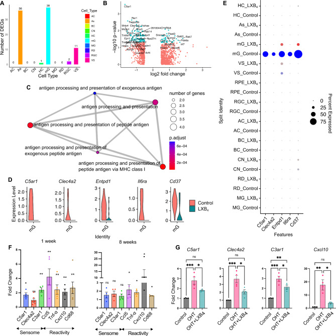
The resident astrocyte-retinal ganglion cell (RGC) lipoxin circuit is impaired during retinal stress, which includes ocular hypertension-induced neuropathy. Lipoxin B produced by homeostatic astrocytes directly acts on RGCs to increase survival and function in ocular hypertension-induced neuropathy. RGC death in the retina and axonal degeneration in the optic nerve are driven by the complex interactions between microglia and macroglia. Whether LXB neuroprotective actions include regulation of other cell types in the retina and/or optic nerve is an important knowledge gap.

METHODS

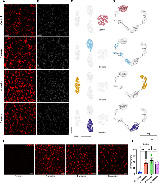
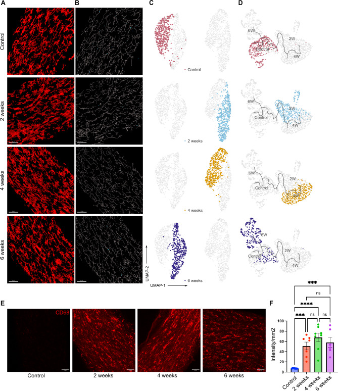
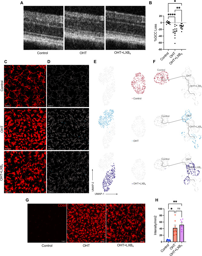
Cellular targets and signaling of LXB in the retina were defined by single-cell RNA sequencing. Retinal neurodegeneration was induced by injecting silicone oil into the anterior chamber of mouse eyes, which induced sustained and stable ocular hypertension. Morphological characterization of microglia populations in the retina and optic nerve was established by MorphOMICs and pseudotime trajectory analyses. The pathways and mechanisms of action of LXB in the optic nerve were investigated using bulk RNA sequencing. Transcriptomics data was validated by qPCR and immunohistochemistry. Differences between experimental groups were assessed by Student's t-test and one-way ANOVA.

RESULTS

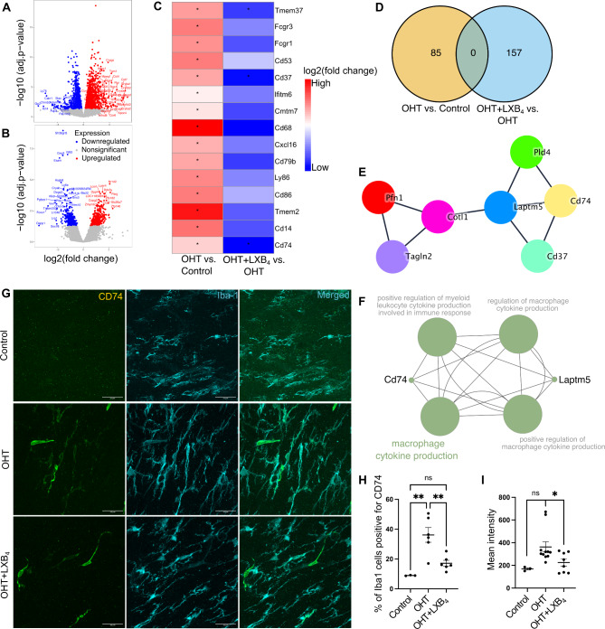
Single-cell transcriptomics identified microglia as a primary target for LXB in the healthy retina. LXB downregulated genes that drive microglia environmental sensing and reactivity responses. Analysis of microglial function revealed that ocular hypertension induced distinct, temporally defined, and dynamic phenotypes in the retina and, unexpectedly, in the distal myelinated optic nerve. Microglial expression of CD74, a marker of disease-associated microglia in the brain, was only induced in a unique population of optic nerve microglia, but not in the retina. Genetic deletion of lipoxin formation correlated with the presence of a CD74 optic nerve microglia population in normotensive eyes, while LXB treatment during ocular hypertension shifted optic nerve microglia toward a homeostatic morphology and non-reactive state and downregulated the expression of CD74. Furthermore, we identified a correlation between CD74 and phospho-phosphoinositide 3-kinases (p-PI3K) expression levels in the optic nerve, which was reduced by LXB treatment.

CONCLUSION

We identified early and dynamic changes in the microglia functional phenotype, reactivity, and induction of a unique CD74 microglia population in the distal optic nerve as key features of ocular hypertension-induced neurodegeneration. Our findings establish microglia regulation as a novel LXB target in the retina and optic nerve. LXB maintenance of a homeostatic optic nerve microglia phenotype and inhibition of a disease-associated phenotype are potential neuroprotective mechanisms for the resident LXB pathway.

摘要

背景

在视网膜应激过程中,包括眼压升高引起的神经病,驻留的星形胶质细胞-视网膜神经节细胞(RGC)脂氧素回路受损。稳态星形胶质细胞产生的脂氧素 B 直接作用于 RGC,增加眼压升高诱导的神经病中的存活和功能。视网膜中 RGC 的死亡和视神经中的轴突变性是由小胶质细胞和大胶质细胞之间的复杂相互作用驱动的。LXB 的神经保护作用是否包括对视网膜和/或视神经中其他细胞类型的调节,这是一个重要的知识空白。

方法

通过单细胞 RNA 测序确定 LXB 在视网膜中的细胞靶标和信号。通过在前房内注射硅油来诱导小鼠眼睛的神经退行性变,从而诱导持续和稳定的眼压升高。通过 MorphOMICs 和拟时轨迹分析建立视网膜和视神经中小胶质细胞群体的形态特征。使用批量 RNA 测序研究 LXB 在视神经中的作用途径和机制。通过 qPCR 和免疫组织化学验证转录组数据。通过 Student's t 检验和单向方差分析评估实验组之间的差异。

结果

单细胞转录组学将小胶质细胞鉴定为健康视网膜中 LXB 的主要靶标。LXB 下调了驱动小胶质细胞环境感应和反应性的基因。小胶质细胞功能分析表明,眼压升高诱导了视网膜和出乎意料的远端有髓视神经中的独特、时间定义和动态表型。CD74 在视神经中仅诱导独特的小胶质细胞群体,但在视网膜中不诱导小胶质细胞的表达,CD74 是大脑中与疾病相关小胶质细胞的标志物。在正常眼压下,脂氧素形成的基因缺失与存在 CD74 视神经小胶质细胞群体相关,而在眼压升高期间,LXB 治疗使视神经小胶质细胞向稳态形态和非反应状态转变,并下调 CD74 的表达。此外,我们发现 CD74 与视神经中磷酸化磷酯酰肌醇 3-激酶(p-PI3K)表达水平之间存在相关性,LXB 治疗降低了这种相关性。

结论

我们确定了小胶质细胞功能表型的早期和动态变化、在远端视神经中诱导独特的 CD74 小胶质细胞群体,这些都是眼压升高诱导的神经退行性变的关键特征。我们的发现确立了小胶质细胞调节作为视网膜和视神经中新型 LXB 靶标。LXB 维持视神经小胶质细胞的稳态表型和抑制疾病相关表型是驻留 LXB 途径的潜在神经保护机制。

https://cdn.ncbi.nlm.nih.gov/pmc/blobs/be5a/11580672/53255f812a22/13024_2024_775_Fig7_HTML.jpg
https://cdn.ncbi.nlm.nih.gov/pmc/blobs/be5a/11580672/6a0ebfec1ad4/13024_2024_775_Fig1_HTML.jpg
https://cdn.ncbi.nlm.nih.gov/pmc/blobs/be5a/11580672/0330361e61df/13024_2024_775_Fig2_HTML.jpg
https://cdn.ncbi.nlm.nih.gov/pmc/blobs/be5a/11580672/fb30a20b40ce/13024_2024_775_Fig3_HTML.jpg
https://cdn.ncbi.nlm.nih.gov/pmc/blobs/be5a/11580672/2a55f72f97d0/13024_2024_775_Fig4_HTML.jpg
https://cdn.ncbi.nlm.nih.gov/pmc/blobs/be5a/11580672/8d38986c67ac/13024_2024_775_Fig5_HTML.jpg
https://cdn.ncbi.nlm.nih.gov/pmc/blobs/be5a/11580672/ab11db96a0cf/13024_2024_775_Fig6_HTML.jpg
https://cdn.ncbi.nlm.nih.gov/pmc/blobs/be5a/11580672/53255f812a22/13024_2024_775_Fig7_HTML.jpg

相似文献

1
Regulation of disease-associated microglia in the optic nerve by lipoxin B and ocular hypertension.脂氧素 B 通过对眼压的调节作用抑制视神经病变中的小胶质细胞。
Mol Neurodegener. 2024 Nov 20;19(1):86. doi: 10.1186/s13024-024-00775-z.
2
Regulation of Diseases-Associated Microglia in the Optic Nerve by Lipoxin B and Ocular Hypertension.脂氧素B与高眼压对视神经中疾病相关小胶质细胞的调控
bioRxiv. 2024 Oct 28:2024.03.18.585452. doi: 10.1101/2024.03.18.585452.
3
Dysregulation of neuroprotective lipoxin pathway in astrocytes in response to cytokines and ocular hypertension​.细胞因子和眼内高压对星形胶质细胞中神经保护脂氧素通路的失调调控。
Acta Neuropathol Commun. 2024 Apr 12;12(1):58. doi: 10.1186/s40478-024-01767-2.
4
Dysregulation of Neuroprotective Lipoxin Pathway in Astrocytes in Response to Cytokines and Ocular Hypertension.星形胶质细胞中神经保护脂氧素通路对细胞因子和高眼压的反应失调。
bioRxiv. 2023 Oct 2:2023.06.22.546157. doi: 10.1101/2023.06.22.546157.
5
IOP induces upregulation of GFAP and MHC-II and microglia reactivity in mice retina contralateral to experimental glaucoma.IOP 诱导实验性青光眼对侧小鼠视网膜 GFAP 和 MHC-II 的上调和小胶质细胞反应性。
J Neuroinflammation. 2012 May 14;9:92. doi: 10.1186/1742-2094-9-92.
6
Astrocyte-derived lipoxins A4 and B4 promote neuroprotection from acute and chronic injury.星形胶质细胞衍生的脂氧素 A4 和 B4 促进急性和慢性损伤的神经保护。
J Clin Invest. 2017 Dec 1;127(12):4403-4414. doi: 10.1172/JCI77398. Epub 2017 Nov 6.
7
Expression and activation of mitogen-activated protein kinases in the optic nerve head in a rat model of ocular hypertension.在眼高压大鼠模型中,视神经头部丝裂原活化蛋白激酶的表达和激活。
Mol Cell Neurosci. 2018 Apr;88:270-291. doi: 10.1016/j.mcn.2018.01.002. Epub 2018 Mar 20.
8
Time-dependent retinal ganglion cell loss, microglial activation and blood-retina-barrier tightness in an acute model of ocular hypertension.高眼压急性模型中视网膜神经节细胞的时间依赖性丢失、小胶质细胞激活及血视网膜屏障紧密性
Exp Eye Res. 2015 Jul;136:59-71. doi: 10.1016/j.exer.2015.05.010. Epub 2015 May 20.
9
Bilateral early activation of retinal microglial cells in a mouse model of unilateral laser-induced experimental ocular hypertension.在单侧激光诱导实验性眼高压小鼠模型中观察到视网膜小胶质细胞的双侧早期激活。
Exp Eye Res. 2018 Jun;171:12-29. doi: 10.1016/j.exer.2018.03.006. Epub 2018 Mar 9.
10
Inhibition of the cysteinyl leukotriene pathways increases survival of RGCs and reduces microglial activation in ocular hypertension.抑制半胱氨酰白三烯途径可增加 RGCs 的存活并减少眼高压中的小胶质细胞激活。
Exp Eye Res. 2021 Dec;213:108806. doi: 10.1016/j.exer.2021.108806. Epub 2021 Oct 27.

引用本文的文献

1
Lipidomic Analysis Reveals Drug-Induced Lipoxins in Glaucoma Treatment.脂质组学分析揭示青光眼治疗中药物诱导的脂氧素
bioRxiv. 2025 Jan 27:2025.01.24.634771. doi: 10.1101/2025.01.24.634771.

本文引用的文献

1
Dysregulation of neuroprotective lipoxin pathway in astrocytes in response to cytokines and ocular hypertension​.细胞因子和眼内高压对星形胶质细胞中神经保护脂氧素通路的失调调控。
Acta Neuropathol Commun. 2024 Apr 12;12(1):58. doi: 10.1186/s40478-024-01767-2.
2
A richer and more diverse future for microglia phenotypes.小胶质细胞表型更丰富多样的未来。
Heliyon. 2023 Mar 21;9(4):e14713. doi: 10.1016/j.heliyon.2023.e14713. eCollection 2023 Apr.
3
Microglia: Friends or Foes in Glaucoma? A Developmental Perspective.小胶质细胞:青光眼的朋友还是敌人?从发育角度看。
Stem Cells Transl Med. 2022 Dec 30;11(12):1210-1218. doi: 10.1093/stcltm/szac077.
4
The STRING database in 2023: protein-protein association networks and functional enrichment analyses for any sequenced genome of interest.2023 年的 STRING 数据库:针对任何感兴趣的测序基因组的蛋白质-蛋白质关联网络和功能富集分析。
Nucleic Acids Res. 2023 Jan 6;51(D1):D638-D646. doi: 10.1093/nar/gkac1000.
5
Microglia depletion exacerbates retinal ganglion cell loss in a mouse model of glaucoma.小胶质细胞耗竭加剧青光眼小鼠模型中的视网膜神经节细胞丢失。
Exp Eye Res. 2022 Dec;225:109273. doi: 10.1016/j.exer.2022.109273. Epub 2022 Oct 4.
6
A tool for mapping microglial morphology, morphOMICs, reveals brain-region and sex-dependent phenotypes.一种用于绘制小胶质细胞形态的工具,morphOMICs,揭示了与大脑区域和性别相关的表型。
Nat Neurosci. 2022 Oct;25(10):1379-1393. doi: 10.1038/s41593-022-01167-6. Epub 2022 Sep 30.
7
Restoration of mitochondria axonal transport by adaptor Disc1 supplementation prevents neurodegeneration and rescues visual function.通过衔接蛋白 Disc1 的补充来恢复线粒体轴突运输可防止神经退行性变并挽救视觉功能。
Cell Rep. 2022 Sep 13;40(11):111324. doi: 10.1016/j.celrep.2022.111324.
8
The PI3K/AKT signalling pathway in inflammation, cell death and glial scar formation after traumatic spinal cord injury: Mechanisms and therapeutic opportunities.创伤性脊髓损伤后炎症、细胞死亡和神经胶质瘢痕形成中的 PI3K/AKT 信号通路:机制和治疗机会。
Cell Prolif. 2022 Sep;55(9):e13275. doi: 10.1111/cpr.13275. Epub 2022 Jun 26.
9
Microglia: Key Players in Retinal Ageing and Neurodegeneration.小胶质细胞:视网膜衰老和神经退行性变的关键参与者。
Front Cell Neurosci. 2022 Mar 17;16:804782. doi: 10.3389/fncel.2022.804782. eCollection 2022.
10
Solving neurodegeneration: common mechanisms and strategies for new treatments.解决神经退行性疾病:新疗法的共同机制和策略。
Mol Neurodegener. 2022 Mar 21;17(1):23. doi: 10.1186/s13024-022-00524-0.