Moulin Chloe, Dvoriantchikova Galina, Bineshfar Niloufar, Swingle Ben, Martinez Gaby, Groso Daniel, Zhang Michelle, Ivanov Dmitry, Pelaez Daniel
Bascom Palmer Eye Institute, University of Miami Miller School of Medicine.
Res Sq. 2024 Nov 2:rs.3.rs-5085599. doi: 10.21203/rs.3.rs-5085599/v1.
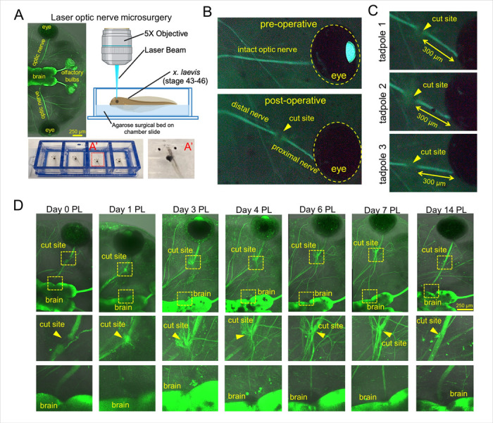
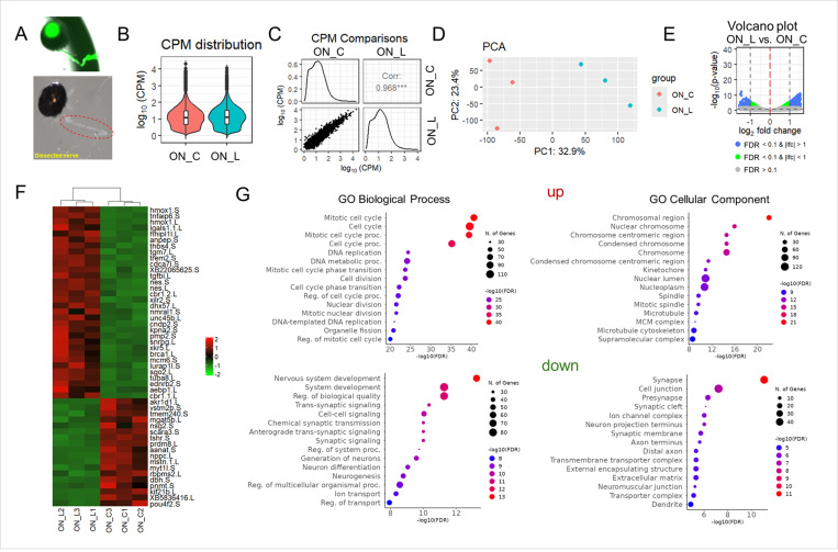
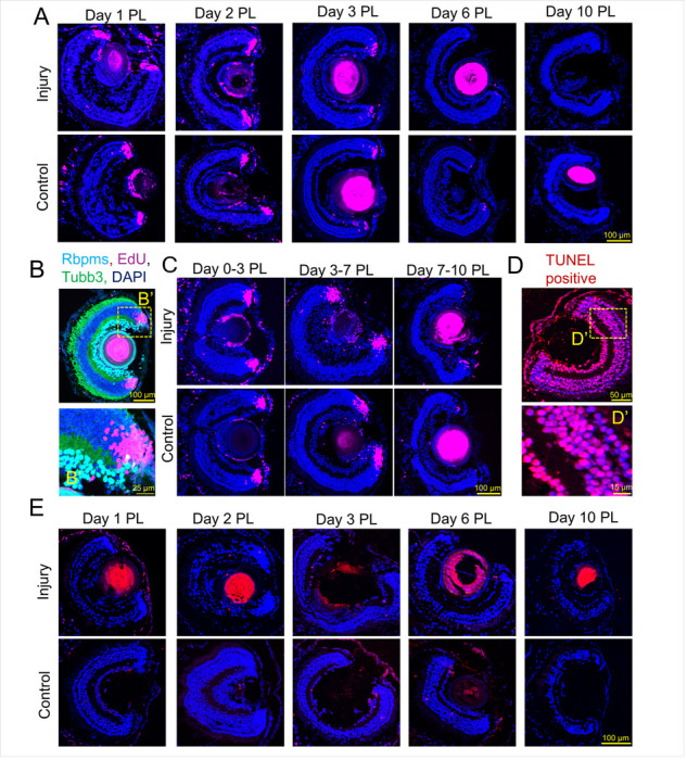
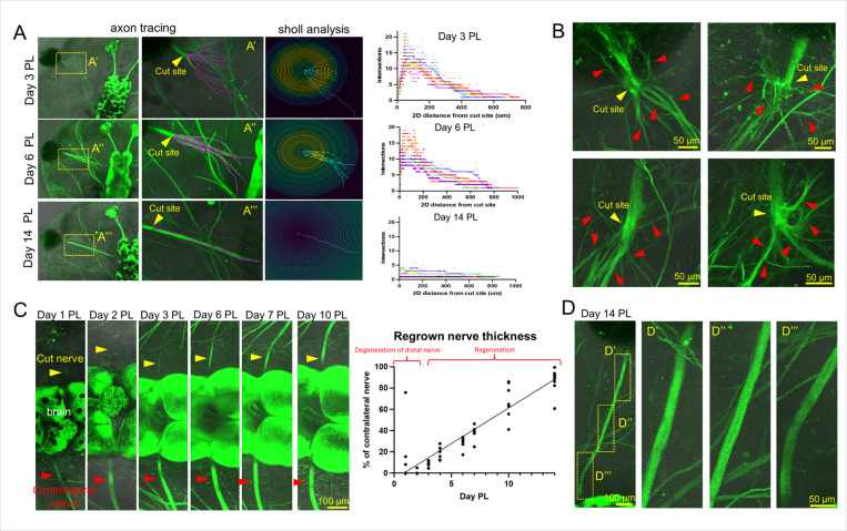
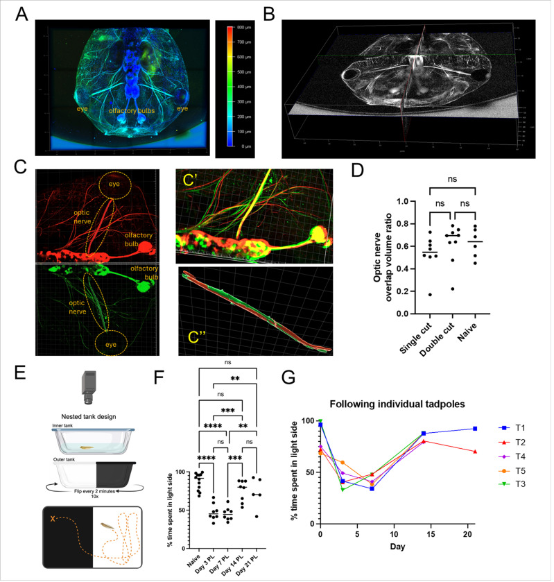
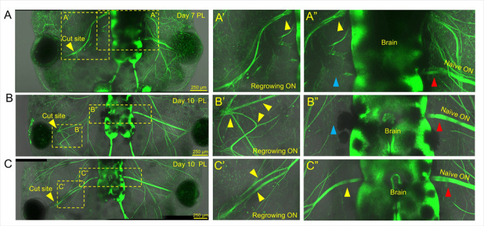
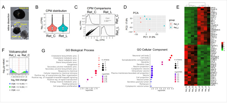
Optic nerve (ON) injury causes blindness in adult mammals as their retinal ganglion cells (RGCs) cannot regenerate axons. However, amphibian RGC axons do not experience the same regenerative failure. Studying the regeneration process of the ON in amphibians holds profound implications for regenerative medicine and human health. Using transgenic tadpoles and laser micro-optics, we developed a reproducible ON transection and regeneration model. Through microscopy, functional testing, TUNEL, EdU assays, and RNA-seq, we characterized the ON injury response and recovery. Our model suggests no chemoattractant gradient exists early in regeneration, with defasciculated axons sprouting in random directions from the globe-proximal cut end. Once individual axons reach the appropriate anatomical insertion point in the brain, their tract is reinforced by other regenerating axons, restoring normal ON morphology. Thus, guidance cues or scaffolding from brain-innervating axons likely support later stages of regeneration. After 14 days, the regenerated ON is morphologically indistinguishable from the naïve ON, and visual function is restored. We found no evidence of RGC death or new RGC formation in the model, suggesting that only pre-existing RGCs are involved in ON regeneration.
视神经(ON)损伤会导致成年哺乳动物失明,因为它们的视网膜神经节细胞(RGCs)无法再生轴突。然而,两栖动物的RGC轴突不会出现同样的再生失败情况。研究两栖动物视神经的再生过程对再生医学和人类健康具有深远意义。我们利用转基因蝌蚪和激光微光学技术,建立了一个可重复的视神经横断和再生模型。通过显微镜检查、功能测试、TUNEL检测、EdU分析和RNA测序,我们对视神经损伤反应和恢复情况进行了表征。我们的模型表明,在再生早期不存在化学引诱剂梯度,脱束的轴突从眼球近端的切断端向随机方向生长。一旦单个轴突到达大脑中合适的解剖插入点,它们的束会被其他再生轴突加强,从而恢复正常的视神经形态。因此,来自支配大脑的轴突的引导线索或支架可能支持再生的后期阶段。14天后,再生的视神经在形态上与未受损的视神经无法区分,视觉功能得以恢复。我们在模型中没有发现RGC死亡或新的RGC形成的证据,这表明只有预先存在的RGC参与了视神经再生。