• 文献检索
  • 文档翻译
  • 深度研究
  • 学术资讯
  • Suppr Zotero 插件Zotero 插件
  • 邀请有礼
  • 套餐&价格
  • 历史记录
应用&插件
Suppr Zotero 插件Zotero 插件浏览器插件Mac 客户端Windows 客户端微信小程序
定价
高级版会员购买积分包购买API积分包
服务
文献检索文档翻译深度研究API 文档MCP 服务
关于我们
关于 Suppr公司介绍联系我们用户协议隐私条款
关注我们

Suppr 超能文献

核心技术专利:CN118964589B侵权必究
粤ICP备2023148730 号-1Suppr @ 2026

文献检索

告别复杂PubMed语法,用中文像聊天一样搜索,搜遍4000万医学文献。AI智能推荐,让科研检索更轻松。

立即免费搜索

文件翻译

保留排版,准确专业,支持PDF/Word/PPT等文件格式,支持 12+语言互译。

免费翻译文档

深度研究

AI帮你快速写综述,25分钟生成高质量综述,智能提取关键信息,辅助科研写作。

立即免费体验

健康乳腺组织的综合单细胞衰老图谱揭示了衰老和癌症共有的表观基因组和转录组特征。

Comprehensive single-cell aging atlas of healthy mammary tissues reveals shared epigenomic and transcriptomic signatures of aging and cancer.

作者信息

Angarola Brittany L, Sharma Siddhartha, Katiyar Neerja, Kang Hyeon Gu, Nehar-Belaid Djamel, Park SungHee, Gott Rachel, Eryilmaz Giray N, LaBarge Mark A, Palucka Karolina, Chuang Jeffrey H, Korstanje Ron, Ucar Duygu, Anczukόw Olga

机构信息

The Jackson Laboratory for Genomic Medicine, Farmington, CT, USA.

The Jackson Laboratory, Bar Harbor, ME, USA.

出版信息

Nat Aging. 2025 Jan;5(1):122-143. doi: 10.1038/s43587-024-00751-8. Epub 2024 Nov 25.

DOI:10.1038/s43587-024-00751-8
PMID:39587369
原文链接:https://pmc.ncbi.nlm.nih.gov/articles/PMC11754115/
Abstract

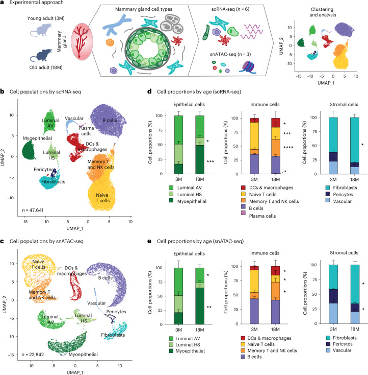
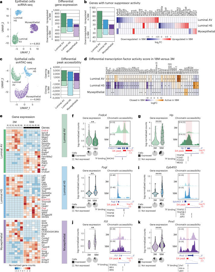
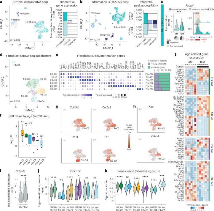
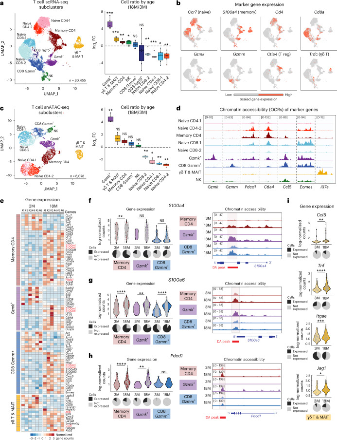
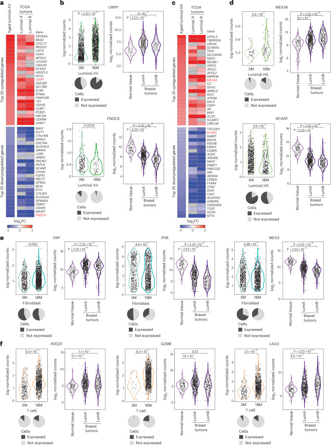
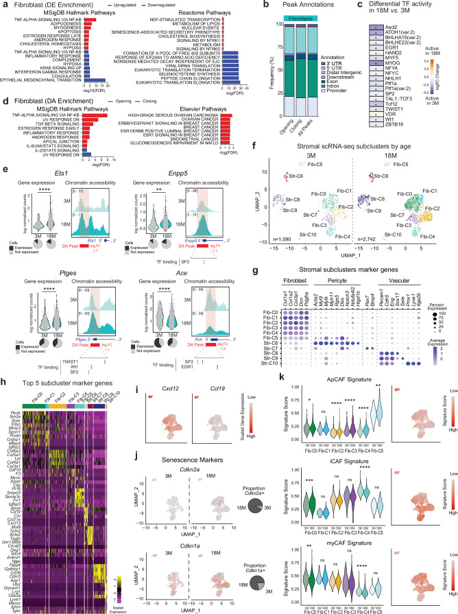
Aging is the greatest risk factor for breast cancer; however, how age-related cellular and molecular events impact cancer initiation is unknown. In this study, we investigated how aging rewires transcriptomic and epigenomic programs of mouse mammary glands at single-cell resolution, yielding a comprehensive resource for aging and cancer biology. Aged epithelial cells exhibit epigenetic and transcriptional changes in metabolic, pro-inflammatory and cancer-associated genes. Aged stromal cells downregulate fibroblast marker genes and upregulate markers of senescence and cancer-associated fibroblasts. Among immune cells, distinct T cell subsets (Gzmk, memory CD4, γδ) and M2-like macrophages expand with age. Spatial transcriptomics reveals co-localization of aged immune and epithelial cells in situ. Lastly, we found transcriptional signatures of aging mammary cells in human breast tumors, suggesting possible links between aging and cancer. Together, these data uncover that epithelial, immune and stromal cells shift in proportions and cell identity, potentially impacting cell plasticity, aged microenvironment and neoplasia risk.

摘要

衰老 是乳腺癌最大的风险因素;然而,与年龄相关的细胞和分子事件如何影响癌症的发生尚不清楚。在本研究中,我们以单细胞分辨率研究了衰老如何重塑小鼠乳腺的转录组和表观基因组程序,为衰老和癌症生物学提供了一个全面的资源。衰老的上皮细胞在代谢、促炎和癌症相关基因中表现出表观遗传和转录变化。衰老的基质细胞下调成纤维细胞标记基因,上调衰老和癌症相关成纤维细胞的标记。在免疫细胞中,不同的T细胞亚群(Gzmk、记忆性CD4、γδ)和M2样巨噬细胞随年龄增长而增加。空间转录组学揭示了衰老的免疫细胞和上皮细胞在原位的共定位。最后,我们在人类乳腺肿瘤中发现了衰老乳腺细胞的转录特征,提示衰老与癌症之间可能存在联系。总之,这些数据揭示了上皮细胞、免疫细胞和基质细胞在比例和细胞特性上的变化,可能影响细胞可塑性、衰老微环境和肿瘤形成风险。

https://cdn.ncbi.nlm.nih.gov/pmc/blobs/89ed/11754115/6735096263f0/43587_2024_751_Fig18_ESM.jpg
https://cdn.ncbi.nlm.nih.gov/pmc/blobs/89ed/11754115/1774e0641f42/43587_2024_751_Fig1_HTML.jpg
https://cdn.ncbi.nlm.nih.gov/pmc/blobs/89ed/11754115/c81624f6f0e6/43587_2024_751_Fig2_HTML.jpg
https://cdn.ncbi.nlm.nih.gov/pmc/blobs/89ed/11754115/9380b79f2e09/43587_2024_751_Fig3_HTML.jpg
https://cdn.ncbi.nlm.nih.gov/pmc/blobs/89ed/11754115/1f2f2cff4cdf/43587_2024_751_Fig4_HTML.jpg
https://cdn.ncbi.nlm.nih.gov/pmc/blobs/89ed/11754115/2dcfed3af821/43587_2024_751_Fig5_HTML.jpg
https://cdn.ncbi.nlm.nih.gov/pmc/blobs/89ed/11754115/6de7d10166cb/43587_2024_751_Fig6_HTML.jpg
https://cdn.ncbi.nlm.nih.gov/pmc/blobs/89ed/11754115/897c0e637369/43587_2024_751_Fig7_HTML.jpg
https://cdn.ncbi.nlm.nih.gov/pmc/blobs/89ed/11754115/a4541b66ca6a/43587_2024_751_Fig8_HTML.jpg
https://cdn.ncbi.nlm.nih.gov/pmc/blobs/89ed/11754115/80da60214e37/43587_2024_751_Fig9_ESM.jpg
https://cdn.ncbi.nlm.nih.gov/pmc/blobs/89ed/11754115/22986f48dec7/43587_2024_751_Fig10_ESM.jpg
https://cdn.ncbi.nlm.nih.gov/pmc/blobs/89ed/11754115/2b2b8b3cd9f5/43587_2024_751_Fig11_ESM.jpg
https://cdn.ncbi.nlm.nih.gov/pmc/blobs/89ed/11754115/a8b93a431565/43587_2024_751_Fig12_ESM.jpg
https://cdn.ncbi.nlm.nih.gov/pmc/blobs/89ed/11754115/bb4fc966ee1b/43587_2024_751_Fig13_ESM.jpg
https://cdn.ncbi.nlm.nih.gov/pmc/blobs/89ed/11754115/41bf79b5ce45/43587_2024_751_Fig14_ESM.jpg
https://cdn.ncbi.nlm.nih.gov/pmc/blobs/89ed/11754115/986db0657817/43587_2024_751_Fig15_ESM.jpg
https://cdn.ncbi.nlm.nih.gov/pmc/blobs/89ed/11754115/316384526b8e/43587_2024_751_Fig16_ESM.jpg
https://cdn.ncbi.nlm.nih.gov/pmc/blobs/89ed/11754115/a9413bcfc4be/43587_2024_751_Fig17_ESM.jpg
https://cdn.ncbi.nlm.nih.gov/pmc/blobs/89ed/11754115/6735096263f0/43587_2024_751_Fig18_ESM.jpg

相似文献

1
Comprehensive single-cell aging atlas of healthy mammary tissues reveals shared epigenomic and transcriptomic signatures of aging and cancer.健康乳腺组织的综合单细胞衰老图谱揭示了衰老和癌症共有的表观基因组和转录组特征。
Nat Aging. 2025 Jan;5(1):122-143. doi: 10.1038/s43587-024-00751-8. Epub 2024 Nov 25.
2
Comprehensive single cell aging atlas of mammary tissues reveals shared epigenomic and transcriptomic signatures of aging and cancer.乳腺组织的综合单细胞衰老图谱揭示了衰老和癌症共有的表观基因组和转录组特征。
bioRxiv. 2023 Oct 23:2023.10.20.563147. doi: 10.1101/2023.10.20.563147.
3
Aging-Associated Alterations in Mammary Epithelia and Stroma Revealed by Single-Cell RNA Sequencing.单细胞 RNA 测序揭示的与衰老相关的乳腺上皮和基质改变。
Cell Rep. 2020 Dec 29;33(13):108566. doi: 10.1016/j.celrep.2020.108566.
4
Integration of microRNA signatures of distinct mammary epithelial cell types with their gene expression and epigenetic portraits.不同乳腺上皮细胞类型的微小RNA特征与其基因表达和表观遗传图谱的整合。
Breast Cancer Res. 2015 Jun 18;17(1):85. doi: 10.1186/s13058-015-0585-0.
5
Consequences of epithelial or stromal TGFβ1 depletion in the mammary gland.上皮或基质 TGFβ1 耗竭对乳腺的影响。
J Mammary Gland Biol Neoplasia. 2011 Jun;16(2):147-55. doi: 10.1007/s10911-011-9218-0. Epub 2011 May 17.
6
Progressive senescence programs induce intrinsic vulnerability to aging-related female breast cancer.衰老相关进程诱导固有脆弱性导致女性乳腺癌。
Nat Commun. 2024 Jun 17;15(1):5154. doi: 10.1038/s41467-024-49106-2.
7
Transcriptome analysis of embryonic mammary cells reveals insights into mammary lineage establishment.胚胎乳腺细胞转录组分析揭示了乳腺谱系建立的见解。
Breast Cancer Res. 2011 Aug 11;13(4):R79. doi: 10.1186/bcr2928.
8
Embryonic mammary signature subsets are activated in Brca1-/- and basal-like breast cancers.胚胎期乳腺特征亚群在Brca1基因敲除小鼠及基底样乳腺癌中被激活。
Breast Cancer Res. 2013 Mar 18;15(2):R25. doi: 10.1186/bcr3403.
9
Characterization of Gene Expression Signatures for the Identification of Cellular Heterogeneity in the Developing Mammary Gland.发育中的乳腺中细胞异质性鉴定的基因表达特征的描述。
J Mammary Gland Biol Neoplasia. 2021 Mar;26(1):43-66. doi: 10.1007/s10911-021-09486-3. Epub 2021 May 14.
10
Single-cell RNA-Seq reveals cell heterogeneity and hierarchy within mouse mammary epithelia.单细胞 RNA 测序揭示了小鼠乳腺上皮细胞内的细胞异质性和层次结构。
J Biol Chem. 2018 Jun 1;293(22):8315-8329. doi: 10.1074/jbc.RA118.002297. Epub 2018 Apr 17.

引用本文的文献

1
The Role of Senescence, its Therapeutic Relevance and Clinical Implications in the Tumor Microenvironment.衰老在肿瘤微环境中的作用、其治疗相关性及临床意义
Theranostics. 2025 Jul 28;15(16):8675-8703. doi: 10.7150/thno.112633. eCollection 2025.
2
Ageing, immune fitness and cancer.衰老、免疫健康与癌症。
Nat Rev Cancer. 2025 Aug 14. doi: 10.1038/s41568-025-00858-z.
3
Beyond polarization: macrophage senescence in immunoregulation and cancer therapy.超越极化:巨噬细胞衰老在免疫调节和癌症治疗中的作用

本文引用的文献

1
Luminal epithelial cells integrate variable responses to aging into stereotypical changes that underlie breast cancer susceptibility.管腔上皮细胞将对衰老的多种反应整合为构成乳腺癌易感性基础的典型变化。
Elife. 2024 Nov 15;13:e95720. doi: 10.7554/eLife.95720.
2
Midkine as a driver of age-related changes and increase in mammary tumorigenesis.Midkine 作为与年龄相关的变化和乳腺肿瘤发生增加的驱动因素。
Cancer Cell. 2024 Nov 11;42(11):1936-1954.e9. doi: 10.1016/j.ccell.2024.09.002. Epub 2024 Oct 3.
3
Senescent CAFs Mediate Immunosuppression and Drive Breast Cancer Progression.
Int J Biol Sci. 2025 Jun 23;21(10):4312-4333. doi: 10.7150/ijbs.115921. eCollection 2025.
4
Plasticity and Functional Heterogeneity of Cancer-Associated Fibroblasts.癌症相关成纤维细胞的可塑性与功能异质性
Cancer Res. 2025 Jul 29. doi: 10.1158/0008-5472.CAN-24-3037.
5
Investigating Transcriptional Age Acceleration in Inflammatory Skin Diseases.炎症性皮肤病中转录年龄加速的研究
JID Innov. 2025 May 30;5(5):100386. doi: 10.1016/j.xjidi.2025.100386. eCollection 2025 Sep.
6
Single-cell analysis of gene regulatory networks in the mammary glands of P4HA1-knockout mice.P4HA1基因敲除小鼠乳腺中基因调控网络的单细胞分析。
PLoS Genet. 2025 Jul 22;21(7):e1011505. doi: 10.1371/journal.pgen.1011505. eCollection 2025 Jul.
7
The Aging Microenvironment as a Determinant of Immune Exclusion and Metastatic Fate in Pancreatic Cancer.衰老微环境作为胰腺癌免疫排斥和转移命运的决定因素
bioRxiv. 2025 May 10:2025.05.08.652966. doi: 10.1101/2025.05.08.652966.
8
Reciprocal immune-epithelial interaction during breast cancer induction.乳腺癌诱发过程中的免疫-上皮相互作用
J Immunother Cancer. 2025 Jun 19;13(6):e011453. doi: 10.1136/jitc-2024-011453.
9
Transforming Cancer Therapy: Unlocking the Potential of Targeting Vascular and Stromal Cells in the Tumor Microenvironment.变革癌症治疗:挖掘肿瘤微环境中靶向血管和基质细胞的潜力
Cancer Res. 2025 Apr 2. doi: 10.1158/0008-5472.CAN-24-4744.
10
Immuno-oncology recapitulates ontogeny: Modern cell and gene therapy for cancer.免疫肿瘤学重现个体发生:癌症的现代细胞和基因疗法。
Mol Ther. 2025 May 7;33(5):2229-2237. doi: 10.1016/j.ymthe.2025.03.042. Epub 2025 Mar 27.
衰老的癌相关成纤维细胞介导免疫抑制并推动乳腺癌进展。
Cancer Discov. 2024 Jul 1;14(7):1302-1323. doi: 10.1158/2159-8290.CD-23-0426.
4
Single Cell Atlas: a single-cell multi-omics human cell encyclopedia.单细胞图谱:单细胞多组学人类细胞百科全书。
Genome Biol. 2024 Apr 19;25(1):104. doi: 10.1186/s13059-024-03246-2.
5
A single-cell atlas enables mapping of homeostatic cellular shifts in the adult human breast.单细胞图谱可绘制成年人大脑乳腺细胞的稳态变化。
Nat Genet. 2024 Apr;56(4):652-662. doi: 10.1038/s41588-024-01688-9. Epub 2024 Mar 28.
6
The cycling and aging mouse female reproductive tract at single-cell resolution.单细胞分辨率下的周期性和衰老的雌性小鼠生殖道。
Cell. 2024 Feb 15;187(4):981-998.e25. doi: 10.1016/j.cell.2024.01.021. Epub 2024 Feb 6.
7
Loss of epigenetic suppression of retrotransposons with oncogenic potential in aging mammary luminal epithelial cells.衰老的乳腺腔上皮细胞中端粒反转录转座子致癌潜能的表观遗传抑制丧失。
Genome Res. 2023 Aug;33(8):1229-1241. doi: 10.1101/gr.277511.122. Epub 2023 Jul 18.
8
A spatially resolved single-cell genomic atlas of the adult human breast.成人乳腺的空间分辨单细胞基因组图谱。
Nature. 2023 Aug;620(7972):181-191. doi: 10.1038/s41586-023-06252-9. Epub 2023 Jun 28.
9
MYC regulates a pan-cancer network of co-expressed oncogenic splicing factors.MYC 调控一个在多种癌症中共同表达的致癌剪接因子的泛癌网络。
Cell Rep. 2022 Nov 22;41(8):111704. doi: 10.1016/j.celrep.2022.111704.
10
Spatial Positioning and Matrix Programs of Cancer-Associated Fibroblasts Promote T-cell Exclusion in Human Lung Tumors.癌症相关成纤维细胞的空间定位和基质程序促进人肺肿瘤中的 T 细胞排斥。
Cancer Discov. 2022 Nov 2;12(11):2606-2625. doi: 10.1158/2159-8290.CD-21-1714.