• 文献检索
  • 文档翻译
  • 深度研究
  • 学术资讯
  • Suppr Zotero 插件Zotero 插件
  • 邀请有礼
  • 套餐&价格
  • 历史记录
应用&插件
Suppr Zotero 插件Zotero 插件浏览器插件Mac 客户端Windows 客户端微信小程序
定价
高级版会员购买积分包购买API积分包
服务
文献检索文档翻译深度研究API 文档MCP 服务
关于我们
关于 Suppr公司介绍联系我们用户协议隐私条款
关注我们

Suppr 超能文献

核心技术专利:CN118964589B侵权必究
粤ICP备2023148730 号-1Suppr @ 2026

文献检索

告别复杂PubMed语法,用中文像聊天一样搜索,搜遍4000万医学文献。AI智能推荐,让科研检索更轻松。

立即免费搜索

文件翻译

保留排版,准确专业,支持PDF/Word/PPT等文件格式,支持 12+语言互译。

免费翻译文档

深度研究

AI帮你快速写综述,25分钟生成高质量综述,智能提取关键信息,辅助科研写作。

立即免费体验

人工智能在急诊科胸部X光片中的诊断性能

Diagnostic Performance of Artificial Intelligence in Chest Radiographs Referred from the Emergency Department.

作者信息

López Alcolea Julia, Fernández Alfonso Ana, Cano Alonso Raquel, Álvarez Vázquez Ana, Díaz Moreno Alejandro, García Castellanos David, Sanabria Greciano Lucía, Hayoun Chawar, Recio Rodríguez Manuel, Andreu Vázquez Cristina, Thuissard Vasallo Israel John, Martínez de Vega Vicente

机构信息

Hospital Universitario QuironSalud Madrid, 28223 Madrid, Spain.

Faculty of Biomedical and Health Science, Universidad Europea de Madrid, 28670 Madrid, Spain.

出版信息

Diagnostics (Basel). 2024 Nov 18;14(22):2592. doi: 10.3390/diagnostics14222592.

DOI:10.3390/diagnostics14222592
PMID:39594258
原文链接:https://pmc.ncbi.nlm.nih.gov/articles/PMC11592727/
Abstract

BACKGROUND

The increasing integration of AI in chest X-ray evaluation holds promise for enhancing diagnostic accuracy and optimizing clinical workflows. However, understanding its performance in real-world clinical settings is essential.

OBJECTIVES

In this study, we evaluated the sensitivity (Se) and specificity (Sp) of an AI-based software (Arterys MICA v29.4.0) alongside a radiology resident in interpreting chest X-rays referred from the emergency department (ED), using a senior radiologist's assessment as the gold standard (GS). We assessed the concordance between the AI system and the resident, noted the frequency of doubtful cases for each category, identified how many were considered positive by the GS, and assessed variables that AI was not trained to detect.

METHODS

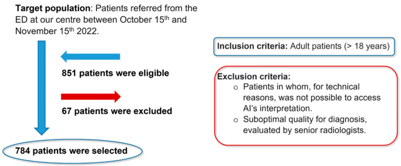
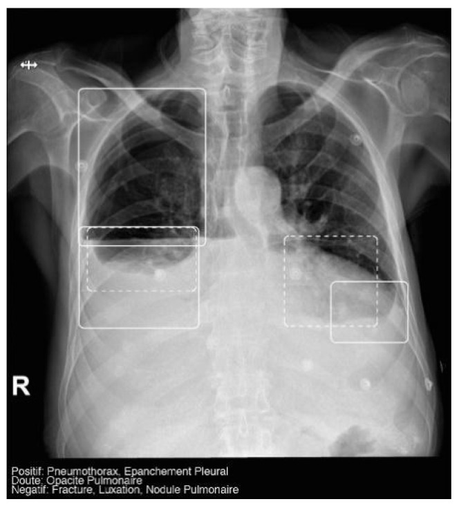
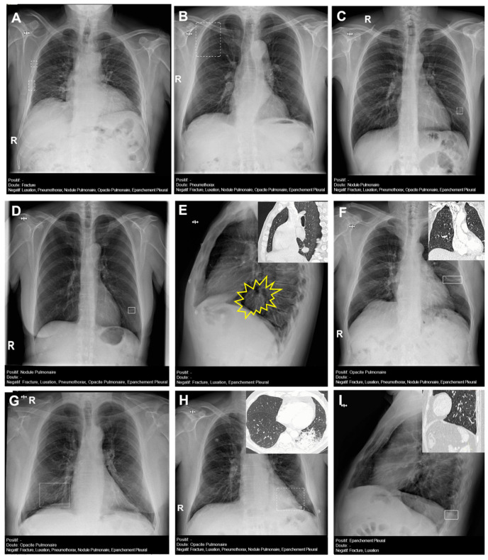
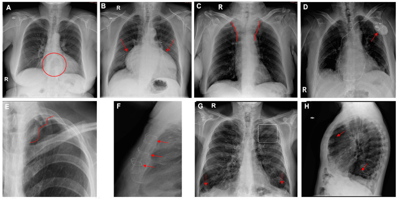
We conducted a retrospective observational study analyzing chest X-rays from a sample of 784 patients referred from the ED at our hospital. The AI system was trained to detect five categorical variables-pulmonary nodule, pulmonary opacity, pleural effusion, pneumothorax, and fracture-and assign each a confidence label ("positive", "doubtful", or "negative").

RESULTS

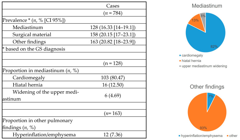
Sensitivity in detecting fractures and pneumothorax was high (100%) for both AI and the resident, moderate for pulmonary opacity (AI = 76%, resident = 71%), and acceptable for pleural effusion (AI = 60%, resident = 67%), with negative predictive values (NPV) above 95% and areas under the curve (AUC) exceeding 0.8. The resident showed moderate sensitivity (75%) for pulmonary nodules, while AI's sensitivity was low (33%). AI assigned a "doubtful" label to some diagnoses, most of which were deemed negative by the GS; the resident expressed doubt less frequently. The Kappa coefficient between the resident and AI was fair (0.3) across most categories, except for pleural effusion, where concordance was moderate (0.5). Our study highlighted additional findings not detected by AI, including 16% prevalence of mediastinal abnormalities, 20% surgical materials, and 20% other pulmonary findings.

CONCLUSIONS

Although AI demonstrated utility in identifying most primary findings-except for pulmonary nodules-its high NPV suggests it may be valuable for screening. Further training of the AI software and broadening its scope to identify additional findings could enhance its detection capabilities and increase its applicability in clinical practice.

摘要

背景

人工智能在胸部X线评估中的日益融合有望提高诊断准确性并优化临床工作流程。然而,了解其在实际临床环境中的表现至关重要。

目的

在本研究中,我们以一位资深放射科医生的评估作为金标准,评估了基于人工智能的软件(Arterys MICA v29.4.0)与放射科住院医师在解读急诊科转诊的胸部X线片时的敏感性(Se)和特异性(Sp)。我们评估了人工智能系统与住院医师之间的一致性,记录了每个类别中可疑病例的频率,确定了金标准认为阳性的病例数量,并评估了人工智能未训练检测的变量。

方法

我们进行了一项回顾性观察研究,分析了我院急诊科转诊的784例患者的胸部X线片样本。人工智能系统经过训练以检测五个分类变量——肺结节、肺部实变、胸腔积液、气胸和骨折——并为每个变量分配一个置信标签(“阳性”、“可疑”或“阴性”)。

结果

人工智能和住院医师检测骨折和气胸的敏感性都很高(100%),检测肺部实变的敏感性中等(人工智能 = 76%,住院医师 = 71%),检测胸腔积液的敏感性尚可(人工智能 = 60%,住院医师 = 67%),阴性预测值(NPV)高于95%,曲线下面积(AUC)超过0.8。住院医师检测肺结节的敏感性中等(75%),而人工智能的敏感性较低(33%)。人工智能为一些诊断分配了“可疑”标签,其中大多数被金标准判定为阴性;住院医师表达怀疑的频率较低。除胸腔积液的一致性为中等(0.5)外,住院医师与人工智能之间的Kappa系数在大多数类别中为一般(0.3)。我们的研究突出了人工智能未检测到的其他发现,包括16%的纵隔异常患病率、20%的手术材料和20%的其他肺部发现。

结论

尽管人工智能在识别大多数主要发现方面显示出效用——除了肺结节——但其高阴性预测值表明它可能对筛查有价值。对人工智能软件进行进一步训练并扩大其识别其他发现的范围,可以提高其检测能力并增加其在临床实践中的适用性。

https://cdn.ncbi.nlm.nih.gov/pmc/blobs/f88a/11592727/9ccb12bd9045/diagnostics-14-02592-g007.jpg
https://cdn.ncbi.nlm.nih.gov/pmc/blobs/f88a/11592727/2436d95f9ee6/diagnostics-14-02592-g001.jpg
https://cdn.ncbi.nlm.nih.gov/pmc/blobs/f88a/11592727/b80e843b75e9/diagnostics-14-02592-g002.jpg
https://cdn.ncbi.nlm.nih.gov/pmc/blobs/f88a/11592727/d87379c5b89c/diagnostics-14-02592-g003.jpg
https://cdn.ncbi.nlm.nih.gov/pmc/blobs/f88a/11592727/3ddbb5783506/diagnostics-14-02592-g004.jpg
https://cdn.ncbi.nlm.nih.gov/pmc/blobs/f88a/11592727/0b753d282a2c/diagnostics-14-02592-g005.jpg
https://cdn.ncbi.nlm.nih.gov/pmc/blobs/f88a/11592727/11485e20a40a/diagnostics-14-02592-g006.jpg
https://cdn.ncbi.nlm.nih.gov/pmc/blobs/f88a/11592727/9ccb12bd9045/diagnostics-14-02592-g007.jpg

相似文献

1
Diagnostic Performance of Artificial Intelligence in Chest Radiographs Referred from the Emergency Department.人工智能在急诊科胸部X光片中的诊断性能
Diagnostics (Basel). 2024 Nov 18;14(22):2592. doi: 10.3390/diagnostics14222592.
2
Diagnostic Performance of an Artificial Intelligence Software for the Evaluation of Bone X-Ray Examinations Referred from the Emergency Department.用于评估急诊科转诊的骨骼X光检查的人工智能软件的诊断性能
Diagnostics (Basel). 2025 Feb 18;15(4):491. doi: 10.3390/diagnostics15040491.
3
Commercially Available Chest Radiograph AI Tools for Detecting Airspace Disease, Pneumothorax, and Pleural Effusion.用于检测气腔疾病、气胸和胸腔积液的商用胸部X光人工智能工具。
Radiology. 2023 Sep;308(3):e231236. doi: 10.1148/radiol.231236.
4
Navigating the Spectrum: Assessing the Concordance of ML-Based AI Findings with Radiology in Chest X-Rays in Clinical Settings.探索全谱:评估临床环境中基于机器学习的人工智能在胸部X光检查结果与放射学结果的一致性。
Healthcare (Basel). 2024 Nov 7;12(22):2225. doi: 10.3390/healthcare12222225.
5
Comparison of Chest Radiograph Interpretations by Artificial Intelligence Algorithm vs Radiology Residents.人工智能算法与放射科住院医师对胸部 X 线片解读的比较。
JAMA Netw Open. 2020 Oct 1;3(10):e2022779. doi: 10.1001/jamanetworkopen.2020.22779.
6
Diagnostic performance of artificial intelligence in interpreting thyroid nodules on ultrasound images: a multicenter retrospective study.人工智能在解读甲状腺结节超声图像中的诊断性能:一项多中心回顾性研究。
Quant Imaging Med Surg. 2024 May 1;14(5):3676-3694. doi: 10.21037/qims-23-1650. Epub 2024 Apr 23.
7
Diagnostic performance of artificial intelligence approved for adults for the interpretation of pediatric chest radiographs.人工智能在成人胸部 X 线片解读方面的诊断性能。
Sci Rep. 2022 Jun 17;12(1):10215. doi: 10.1038/s41598-022-14519-w.
8
Using AI to Improve Radiologist Performance in Detection of Abnormalities on Chest Radiographs.利用人工智能提高放射科医生在胸部X光片上检测异常的表现。
Radiology. 2023 Dec;309(3):e230860. doi: 10.1148/radiol.230860.
9
An Artificial Intelligence-Based Chest X-ray Model on Human Nodule Detection Accuracy From a Multicenter Study.基于人工智能的多中心研究中用于检测人类结节的胸部 X 射线模型。
JAMA Netw Open. 2021 Dec 1;4(12):e2141096. doi: 10.1001/jamanetworkopen.2021.41096.
10
Validation of a Deep Learning Algorithm for the Detection of Malignant Pulmonary Nodules in Chest Radiographs.深度学习算法在胸部 X 光片中检测恶性肺结节的验证。
JAMA Netw Open. 2020 Sep 1;3(9):e2017135. doi: 10.1001/jamanetworkopen.2020.17135.

引用本文的文献

1
Development of an AI model for pneumothorax imaging: Dataset and model optimization strategies for real-world deployment.用于气胸成像的人工智能模型开发:面向实际应用的数据集与模型优化策略
Eur J Radiol Open. 2025 Jun 10;14:100664. doi: 10.1016/j.ejro.2025.100664. eCollection 2025 Jun.
2
Mapping artificial intelligence models in emergency medicine: A scoping review on artificial intelligence performance in emergency care and education.绘制急诊医学中的人工智能模型:关于人工智能在急诊护理和教育中表现的范围综述。
Turk J Emerg Med. 2025 Apr 1;25(2):67-91. doi: 10.4103/tjem.tjem_45_25. eCollection 2025 Apr-Jun.
3
Diagnostic Performance of an Artificial Intelligence Software for the Evaluation of Bone X-Ray Examinations Referred from the Emergency Department.
用于评估急诊科转诊的骨骼X光检查的人工智能软件的诊断性能
Diagnostics (Basel). 2025 Feb 18;15(4):491. doi: 10.3390/diagnostics15040491.