Mathieu Marie-Christine, Suciu Voichita, Tanguy Marie-Laure, Ben Romdhane Neila Ines, Moalla Salma, Harguem-Zayani Sana, Barbe Remy, Balleyguier Corinne, Conversano Angelica, Abbaci Muriel
Department of Medical Biology and Pathology, Gustave Roussy, Université Paris-Saclay, 94805 Villejuif, France.
Surgery and Pathology Photonic Imaging Group, Gustave Roussy, 94805 Villejuif, France.
Life (Basel). 2024 Oct 28;14(11):1384. doi: 10.3390/life14111384.
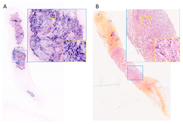
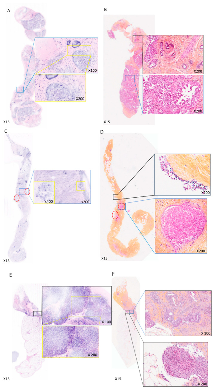
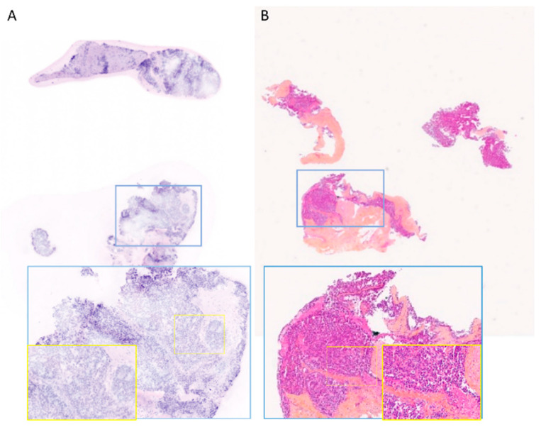
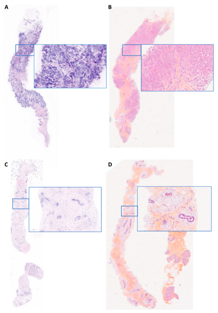
In the one-stop breast clinic setting, breast cytology traditionally provides immediate diagnosis of carcinoma. Fluorescence confocal microscopy (FCM) is an emerging optical technique enabling ex vivo analysis of breast biopsies in real-time. This study represents the first proof of concept for integrating FCM imaging into the routine workflow of breast core needle biopsies (CNB) at Gustave Roussy's one-stop breast clinic.
Fifty women with breast masses underwent consecutive enrollment. Biopsies were stained with acridine orange and fast green, followed by imaging using the Vivascope 2500M-G4 (FCM). Interpretation was conducted by two pathologists in real time (PT1) or postoperatively (PT2). Concordance with definitive histology, the duration of the FCM protocol, and its impact on conventional histopathology, immunohistochemistry, and FISH analyses were evaluated.
In our study of 50 biopsies, a concordant diagnosis of malignancy was performed using FCM on the malignant cases at definitive histology in 93.5% (29/31 cases) and in 90.3% (28/31 cases) according to PT1 and PT2, respectively. When the FCM suspicious cases were added, FCM identified 100% (31/31 cases) and 96.7% (30/31 cases) of the malignant cases according to PT1 and PT2, respectively. A notable false positive case was identified as a complex sclerosing lesion. The median time for sample preparation (including tissue reception) was 5 min, while the median time for imaging acquisition with interpretation was 3 min for PT1, but 1 min required for interpretation alone by PT2. Histopathological alterations were not more prevalent in FCM-imaged biopsies compared to conventionally treated biopsies. The immunophenotyping and molecular assessment of tissue were preserved after FCM protocol.
FCM shows promise as a new histological method for the immediate diagnosis of breast carcinoma on core needle biopsies in a one-stop clinic setting, while also preserving tissue specimens for final histology.
在一站式乳腺诊所环境中,乳腺细胞学传统上可对癌进行即时诊断。荧光共聚焦显微镜(FCM)是一种新兴的光学技术,能够对乳腺活检组织进行实时离体分析。本研究首次证明了将FCM成像整合到古斯塔夫·鲁西一站式乳腺诊所乳腺粗针活检(CNB)常规工作流程中的概念。
连续纳入50例有乳腺肿块的女性。活检组织用吖啶橙和固绿染色,然后使用Vivascope 2500M - G4(FCM)进行成像。由两名病理学家实时(PT1)或术后(PT2)进行解读。评估与最终组织学结果的一致性、FCM方案的持续时间及其对传统组织病理学、免疫组织化学和FISH分析的影响。
在我们对50例活检组织的研究中,根据PT1和PT2,在最终组织学确诊为恶性的病例中,FCM对恶性病例的诊断一致性分别为93.5%(29/31例)和90.3%(28/31例)。当加入FCM可疑病例时,根据PT1和PT2,FCM分别识别出100%(31/31例)和96.7%(30/31例)的恶性病例。有1例假阳性病例被确定为复杂性硬化性病变。样本制备(包括组织接收)的中位时间为5分钟,而PT1进行成像采集并解读的中位时间为3分钟,PT2单独解读仅需1分钟。与传统处理的活检组织相比,FCM成像的活检组织中组织病理学改变并不更普遍。FCM方案后组织的免疫表型分析和分子评估得以保留。
FCM有望成为一种新的组织学方法,用于在一站式诊所环境中对乳腺粗针活检组织即时诊断乳腺癌,同时还能保留组织标本用于最终组织学检查。