Kapic Ammar, Zaman Khadiza, Nguyen Vien, Prokai-Tatrai Katalin, Prokai Laszlo
Department of Pharmacology and Neuroscience, University of North Texas Health Science Center, Fort Worth, TX 76107, USA.
Pharmaceuticals (Basel). 2024 Nov 9;17(11):1508. doi: 10.3390/ph17111508.
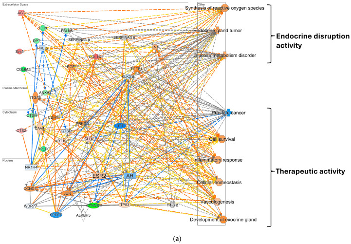
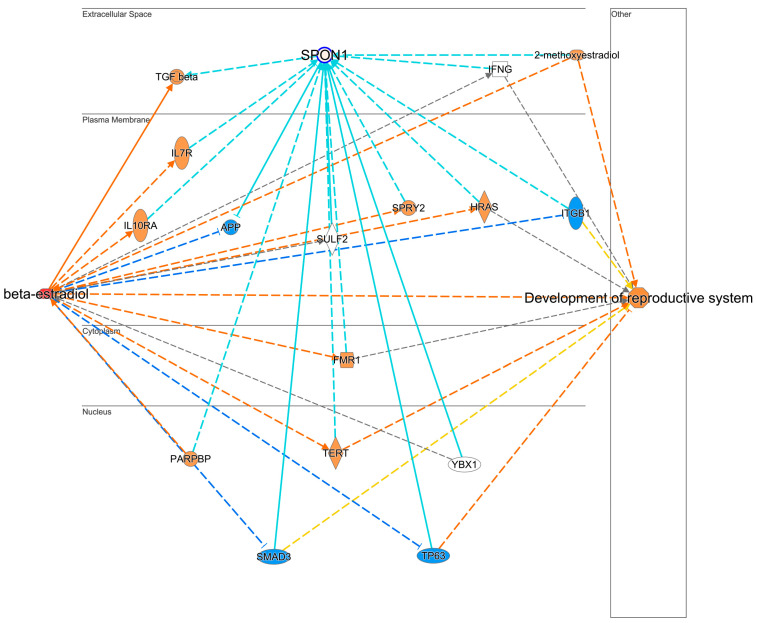
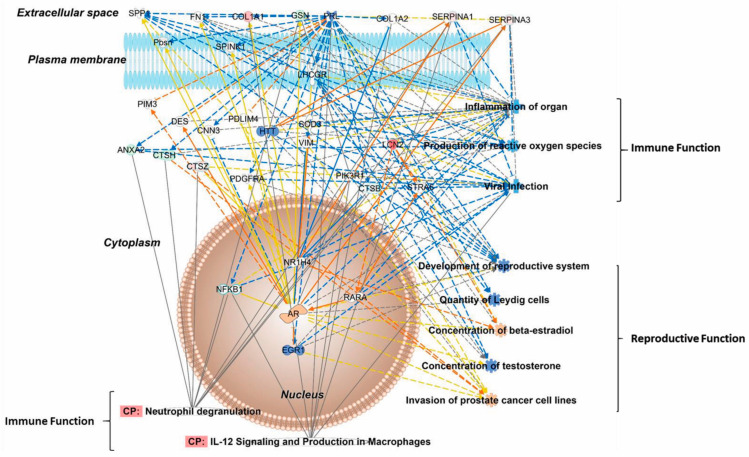
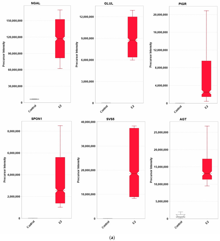
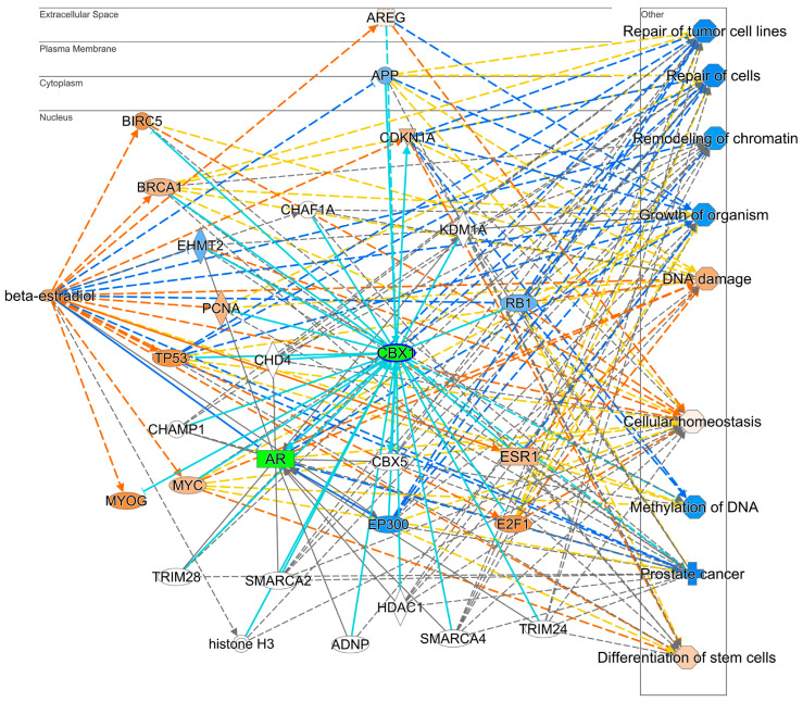
Although estrogenic compounds promise therapeutic potential in treating various conditions, concerns regarding their endocrine-disrupting effects have been raised. Current methodologies for screening estrogenicity in rodent models are limited to the female-specific uterotrophic bioassay. Studies have reported enlargement of the seminal vesicles in orchiectomized males treated with estrogens. However, identifying estrogenicity strictly through changes in wet weights is uninformative regarding the molecular mechanisms of these agents. Therefore, protein-based biomarkers can complement and improve the sensitivity of weight-based assessments. To this end, we present a discovery-driven proteomic analysis of 17β-estradiol's effects on the seminal vesicles. We treated orchidectomized mice with the hormone for five days and used the vehicle-treated group as a control. Seminal vesicles were analyzed by shotgun approach using data-dependent nanoflow liquid chromatography-tandem mass spectrometry and label-free quantification. Proteins found to be differentially expressed between the two groups were processed through a bioinformatics pipeline focusing on pathway analyses and assembly of protein interaction networks. Out of 668 identified proteins that passed rigorous validation criteria, 133 were regulated significantly by 17β-estradiol. Ingenuity Pathway Analysis linked them to several hormone-affected pathways, including those associated with immune function such as neutrophil degranulation. The altered protein interaction networks were also related to functions including endocrine disruption, abnormal metabolism, and therapeutic effects. We identified several potential biomarkers for estrogenicity in mouse seminal vesicles, many of them not previously linked with exogenous 17β-estradiol exposure.
尽管雌激素化合物在治疗各种病症方面具有治疗潜力,但人们对其内分泌干扰作用表示担忧。目前在啮齿动物模型中筛选雌激素活性的方法仅限于雌性特异性子宫增重生物测定法。有研究报告称,接受雌激素治疗的去势雄性动物的精囊会增大。然而,仅通过湿重变化严格鉴定雌激素活性并不能提供有关这些药物分子机制的信息。因此,基于蛋白质的生物标志物可以补充并提高基于重量评估的灵敏度。为此,我们对17β-雌二醇对精囊的作用进行了探索性蛋白质组学分析。我们用该激素对去势小鼠进行了五天的治疗,并将溶剂处理组作为对照。使用数据依赖型纳流液相色谱-串联质谱法和无标记定量法,通过鸟枪法对精囊进行分析。对两组之间差异表达的蛋白质进行生物信息学分析,重点是通路分析和蛋白质相互作用网络的构建。在668种经过严格验证标准鉴定的蛋白质中,有133种受到17β-雌二醇的显著调节。通路分析软件(IPA)将它们与几种受激素影响的通路联系起来,包括与免疫功能相关的通路,如中性粒细胞脱颗粒。改变的蛋白质相互作用网络也与内分泌干扰、异常代谢和治疗效果等功能有关。我们在小鼠精囊中鉴定出了几种雌激素活性的潜在生物标志物,其中许多以前与外源性17β-雌二醇暴露无关。